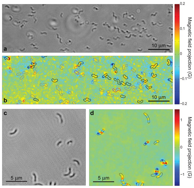
Figure 2. Wide-field optical and magnetic images of magnetotactic bacteria.
a, Bright-field optical image of MTB adhered to the diamond surface while immersed in PBS. b, Image of magnetic field projection along the [111] crystallographic axis in the diamond for the same region as a, determined from NV ODMR. Superimposed outlines indicate MTB locations determined from a. Outline colours indicate results of the live-dead assay performed after measuring the magnetic field (black for living, red for dead, and grey for indeterminate). c, Bright-field image of dried MTB on the diamond chip. d, Image of magnetic field projection along [111] for the same region, with outlines indicating MTB locations determined from c.

