Figure 2.
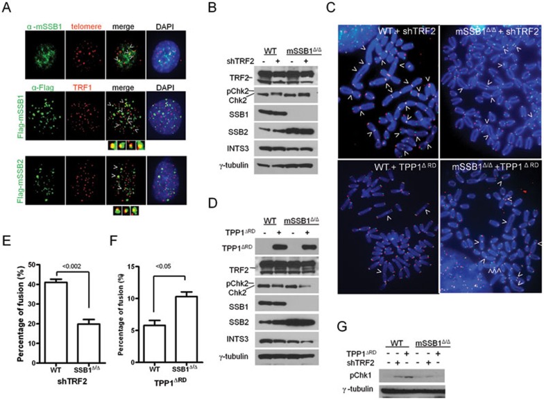
mSSB1 and mSSB2 protect telomeres. (A) Endogenous mSSB1, Flag-mSSB1 and Flag-mSSB2 colocalized with the telomeric PNA probe (telomere: Cy3-OO-(CCCTAA)4 (red)) or the telomere binding protein TRF1 on a subset of telomeres. Colocalized foci are displayed in greater detail in magnified images for Flag-mSSB1 and 2. (B) Immunoblot detection of TRF2, p-Chk2, mSSB1, mSSB2 and INTS3 in MEFs of the indicated genotypes untreated or treated with shRNA against TRF2 for 5 days. γ-tubulin is used as a loading control. (C) Examples of chromosome aberrations in metaphase spreads using Cy3-OO-(CCCTAA)4 (red) and FAM-OO-(TTAGGG)4 (green) telomere peptide nucleic acid probes. Arrows point to sites of chromosome fusions. Not all fusion sites are indicated. (D) Immunoblot detection of TPP1ΔRD, TRF2, pChk2, mSSB1, mSSB2 and INTS3 in MEFs of the indicated genotypes untreated or treated with TPP1ΔRD. γ-tubulin is used as a loading control. (E) Quantification of total telomere fusions in MEFs either untreated or treated with shRNA against TRF2. Two independent cell lines were analyzed, and a minimum of 35 metaphases and 1 500 chromosomes were scored per cell line. Mean values were derived from two independent cell lines. The one-tailed t-test was used to calculate statistical significance. Error bars: s.e.m. (F) Quantification of total telomere fusions in MEFs either untreated or treated with TPP1ΔRD and analyzed as in D. Error bars: s.e.m. (G) Immunoblot detection of p-Chk1 in WT or mSSB1Δ/Δ MEFs untreated or treated with shRNA against TRF2 or expressing TPP1ΔRD. γ-tubulin was used as a loading control.

