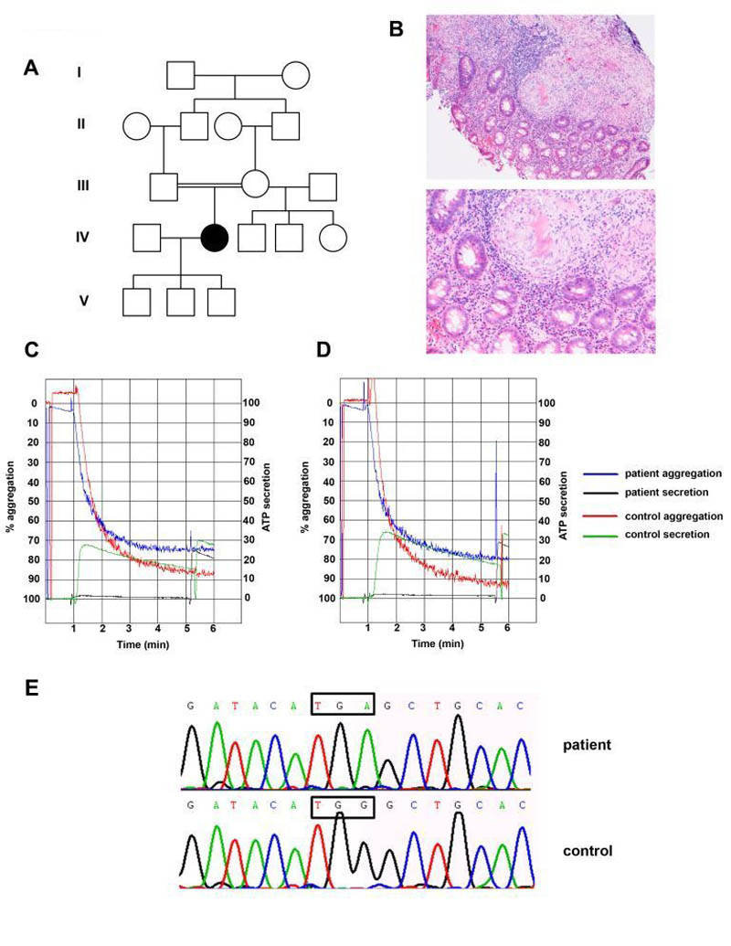
Figure 1. Identification of the second HPS7 mutation in a patient with Hermansky-Pudlak syndrome presenting late in life.
(Panel A) Pedigree of a consanguineous family with Hermansky-Pudlak syndrome. The affected individual is represented by a solid symbol.
(Panel B) Images of a colonic biopsy from the patient in low power (upper panel) and high power (lower panel). Both images show inflammatory infiltrates and granulomata with numerous giant cells, alongside normal bowel mucinous glands. No caseous necrosis is seen, and special stains showed no evidence of micro-organisms (including mycobacteria).
(Panels C and D) Absence of secretion (black trace) to high doses of PAR-1 peptide 100μM (Panel C) and PAR-4 peptide 500μM (Panel D) in this patient. Left sided Y axis depicts percentage aggregation, and right sided Y axis represents platelet ATP secretion assessed using Chronolume®. 1.6nmol of ATP standard were added to each cuvette in order to calculate absolute secretion and secretion normalised to platelet count in PRP for both patient and control.
(Panel E) Identification of a homozygous single base substitution (c.177 G>A) in Dysbindin leading to a premature stop codon (p.Trp59Stop). Sanger sequencing showing wild-type and mutant DTNBP1 sequence traces. The position of the mutation is indicated by the boxed regions.

