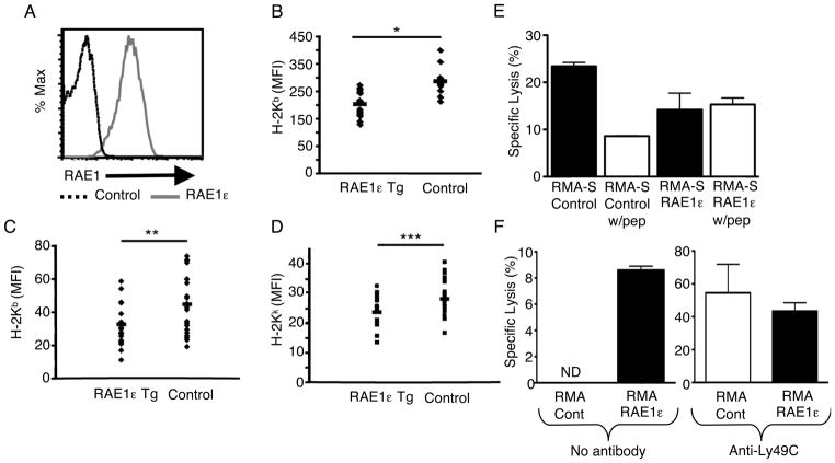
FIGURE 2. Transgenic expression of RAE1ε in vivo results in decreased MHC class I surface expression and increased NK-cell lysis.
(A) PBMCs from RAE1ε transgenic and non-transgenic (control) mice were stained with a RAE1-specific antibody and analyzed by flow cytometry. The data shown are representative of greater than twenty individual mice. (B) PBMCs from RAE1ε transgenic (n=15) and non-transgenic (n=11) mice were stained with a H-2Kb-specific antibody and analyzed by flow cytometry. (C and D) RAE1 transgenic mice were bred to B10.BR mice, and PBMCs from RAE1ε transgenic (n=18) and non-transgenic (n=21) mice were stained with H-2Kb and H-2Kk-specific antibodies and analyzed by flow cytometry. (E and F) NKG2D KO NK-cell lysis (E) against RMA-S/control and RMA-S/RAE1ε cells in the presence or absence of peptide (10μM), or (F) against RMA/control and RMA/RAE1ε, in the presence or absence of anti-Ly49C Fab fragments (200μg/ml), was measured with a standard chromium release assay at an E:T ratio of 30:1. The results are shown as mean + SD of 3 replicates and are representative of two independent experiments. *p=0.03 in a two-sided student’s t test. **p=0.02 in a two-sided student’s t test ***p=0.01 in a two-sided student’s t test. ND: Not detected.

