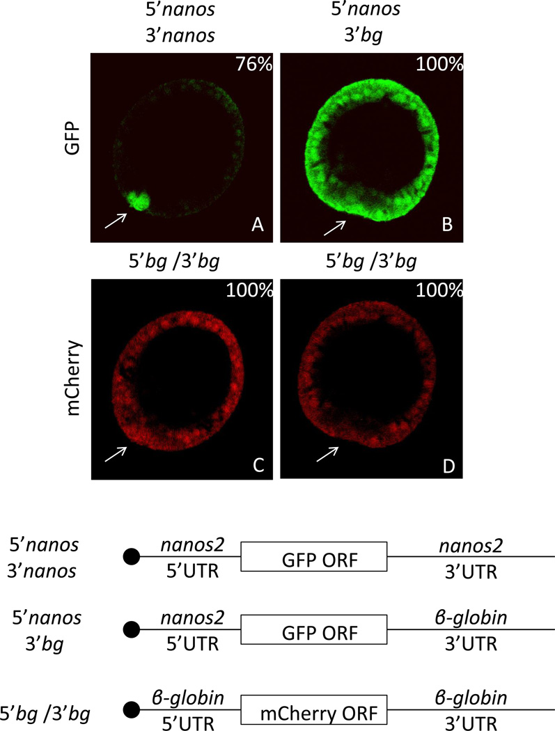
Figure 1.
Sp nanos2 3’UTR is sufficient for the selective protein accumulation in the small micromeres. Synthetic mRNA containing the GFP open reading frame flanked by (A) Sp nanos2 5’ and 3’UTRs or (B) Sp nanos2 5’UTR and Xenopus β-globin 3’UTR were co-injected with mCherry mRNA (C and D) containing Xenopus β-globin 5’ and 3’UTRs in Sp fertilized eggs. GFP (green) and mCherry (red) fluorescence were assayed in the same embryos 24 hours post-fertilization at mesenchyme blastula: A and C represent the same embryo, B and D represent another one. For GFP images, A and B were obtained using the same settings (laser intensity, pin-hole opening). For mCherry images, C and D were also taken using the same settings. bg indicates β-globin UTRs. The arrows are pointing toward the small micromeres. The blastula are presented in the same orientation in the subsequent figures. Approximately one hundred blastulas were visualized after injection of each construct, the corresponding percentages of representative embryos are indicated in the right corner. Cartoons of the injected RNAs are presented at the bottom (the black circle representing the m7GTP cap).

