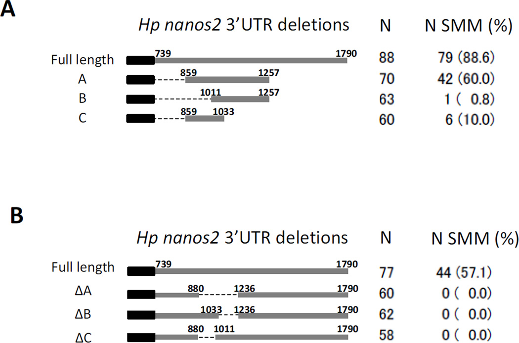
Figure 3.
The nucleotides localized between the position 880 and 1236 of Hp nanos2 3’UTR are essential for protein enrichment in the small micromeres. Synthetic mRNAs were made using Hp nanos2 3’UTR and the GFP ORF followed by different deletions of Hp nanos2 3’UTR (A and B). These RNAs were injected in Hp fertilized eggs, and the numbers of injected embryos having a GFP signal enriched in the small micromeres at the blastula stage was monitored under the fluorescence microscope. N indicates the number of injected embryos used for each RNA construct. N sMic indicates the number of injected embryos having protein enrichment in the small micromeres, the corresponding percentages are represented in parentheses (%). The graphs represent the percentage of injected embryos showing protein enrichment within the small micromeres for each RNA.

