
Figure 3.
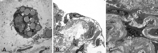
Transmission electron microscopy images of (A) CD133 cells labeled with Dil-acLDL-l that were injected into the lumen of a pulmonary artery. (B) After 4 days incubation, CD133 labeled cells, identified by large cytoplasmic vesicles, differentiated into endothelial cells at the lumen surface. (C) CD133 cells infiltrating the intima acquired a smooth muscle cell-like phenotype.