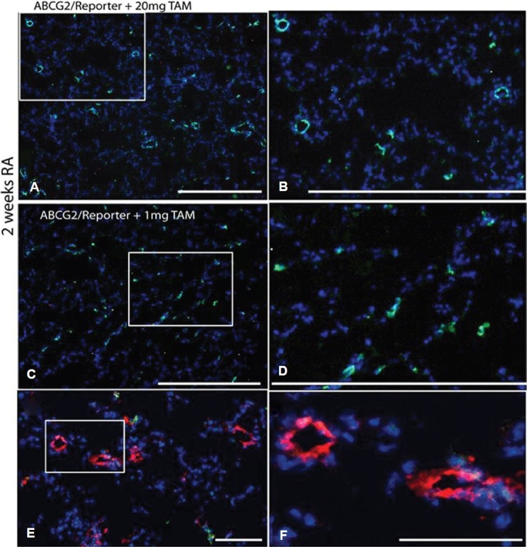
Figure 1.

Optimization of lung MSC targeted recombination in vivo. ABCG2 mouse lung 2 weeks post induction using 20 mg or 1 mg of tamoxifen. Recombination results in the appearance of membrane eGFP expression. (A and B) 20 mg dose tamoxifen labels both vascular EC and mesenchymal cells. (C and D) 1 mg tamoxifen induction labels predominantly mesenchymal cells. Scale bars = 500 μM. (E and F) Factor VIII labeling of vascular endothelium (red) demonstrates lack of detectable ABCG2-positive endothelial cell with low-dose tamoxifen. Scale bars = 50 μM.
