
Figure 4.
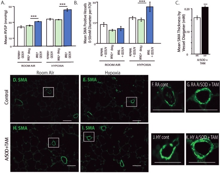
Functional consequences of EC-SOD knockout in lung MSCs include increased muscularization with exacerbated PAH. Physiologic parameters were analyzed to detect how remodeling by MSCs affected PAH. Comparisons are presented between vehicle controls, floxed sod3 + tamoxifen controls and tamoxifen-induced A/SOD mice. In all experiments, 2 weeks after induction, mice were exposed to either ambient air pressure or hypobaric hypoxia for 5 weeks. (A) RVSP was measured via a pressure transducer inserted into the right cardiac ventricle, n = 10, 8, 10, 8, 8, 11. Analysis of the lung tissue was performed to measure histological endpoints associated with MSC remodeling and PAH. (B) Quantitation of muscularization was performed by counting smooth muscle alpha actin (SMA) positive vessels per field of view in paraffin-stained lung sections. Differences were evident in the 0-50 μM diameter vessels; n = 5, 9, 8, 8, 8, 8. (C) The thickness of SMA-positive vessels under 30 μM was measured as a function of vessel diameter; n = 4, 4. (D-K) Representative fluorescent micrographs of SMA (green) stained paraffin lung tissue sections from control and induced (A/SOD + TAM) mice exposed to room air or hypoxia. Enlarged panels of boxed vessels left to right: SMA RA control, RA A/SOD + TAM, hypoxia control, hypoxia A/SOD + TAM; H and E hypoxia A/SOD + TAM. Scale bars = 25 μM.