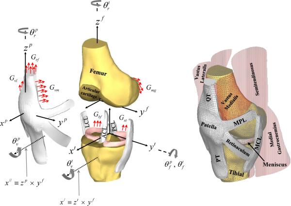
Figure 1.
An anterior-medial view of the three dimensional finite element model of the knee shows the corresponding soft tissues, muscles and articular surfaces acting on the bones. In the figure, ACL and PCL are the anterior and posterior cruciate ligaments, MCL and LCL are the medial and lateral collateral ligaments, MPL and LPL (not shown) are the medial and lateral patello-femoral ligaments, and PT and QT are the patellar and quadriceps tendons, respectively. In all three coordinate systems (patellar, tibial and femoral), the medial (y), anterior (x), and superior (z) directions were chosen to be positive. The femoral and tibial z-axes were in the direction of the long axis of both bones. The patellar z-axis was along the line connecting the center of the patellar coordinates with the lowest point on the apex of the patella. The three dimensional sequential rotations defining TF and PF joint motions shown in the figure, have been described in detail elsewhere (Dhaher and Khan, 2002). For both joints, the first rotation is through an angle about the femoral medio-lateral axis (yf -axis) followed by rotations about the tibial and patellar zaxis (zt and zp) through and respectively. Finally, the last rotation is about an anterior/posterior-floating axis (χ| and χ∥), through and respectively. Finally, Grf, Gvm, Gvl, Gbf, Gmg, Glg, and Gst represent the net muscle force distributed across the common nodes of the muscle-bone junctions.

