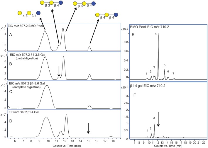
Fig. 3.
(A) EIC of m/z 507.2 from the BMO pool showing four isomers with their respective structures labeled. (B) EIC of m/z 507.2 at the half-way point of digestion with β1-3,6 galactosidase. (C) EIC of m/z 507.2 after full-time digestion with β1-3,6 galactosidase. (D) EIC of m/z 507.2 after digestion with β1-4 galactosidase. (E) EIC of m/z 710.2 from the BMO pool showing seven isomers. (F) EIC of m/z 710.2 after digestion with β1-4 galactosidase.

