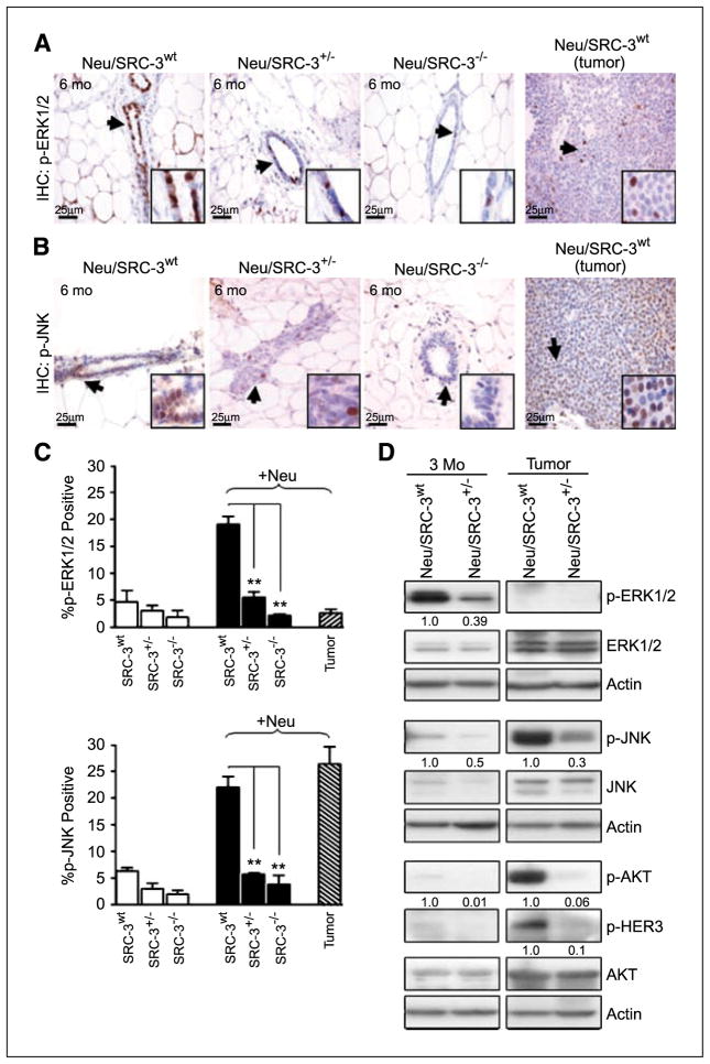
Figure 6.
Lower SRC-3 levels alter Neu-mediated signaling in Neu-induced mammary gland preneoplasia and tumorigenesis. Representative immunohistochemical staining of p-ERK1/2 (A) and p-JNK (B) in mammary gland tissue sections (6 mo of age) and representative Neu-induced mammary tumor sections. Arrows and insets indicate positive staining in mammary epithelial cells (magnification, 40×). C, bar charts represent the quantification of p-ERK1/2 and p-JNK immunohistochemical staining; 100 cells were counted per field, and 10 fields were counted per mouse (magnification, 40×). Columns, mean of three independent experiments (n = 3 mice from each indicated genotype). **, P < 0.001, one-way ANOVA. D, representative Western blot analysis of p-ERK1/2, p-JNK, p-AKT, and p-HER3 and their respective total protein expression levels from whole cell extracts of cultured nontumorigenic mammary epithelial cells and tumor cells. Densitometry values represent p-ERK1/2, p-JNK, p-AKT, and p-HER3 levels normalized to total actin. Western blot results represent data from at least two mice from each genotype.

