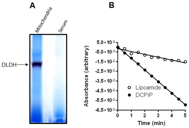
Fig. 4.
Analysis of serum DLDH diaphorase activity and reverse activity. (A) In-gel diaphorase activity staining of serum and mitochondrial DLDH. (B) Spectrophotometric assay of reverse activity using lipoamide (open circle) as the substrate and of diaphorase activity using DCPIP (filled circle) as the electron acceptor. NADH was used as the electron donor in both assays that contained 5 mg serum proteins.

