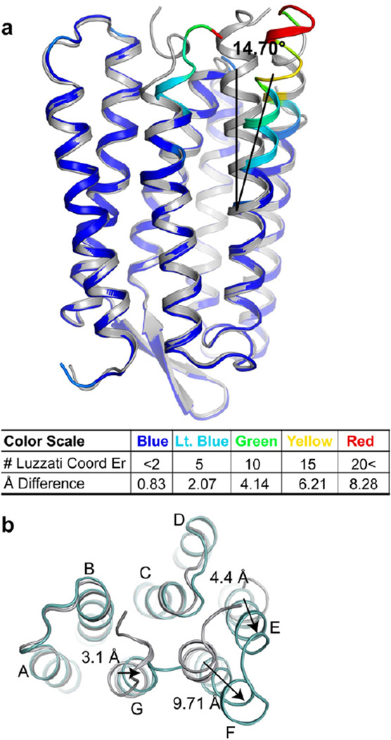
Figure 1. Triple mutant bR structure.
Alignments of the triple mutant (colored) to the wild-type ground state structure (silver) are shown along the membrane plane (panel A) and from the cytoplasmic surface. (a) The color scale represents multiples of the Luzzatti coordinate error illustrating significance of conformational change. Helix F tilts outwards from the ground state structure by 14.70° ± 1.48°. (b) Movement of helix F end relative to the ground state as viewed from the cytoplasmic surface. Arrows indicate direction of movement relative to the wild-type ground state starting point. See also Figure S1.

