
Figure 5.
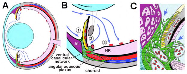
Model of aqueous humor dynamics in the zebrafish eye. (A) Overview showing the vectorial flow of aqueous humor (blue arrows) from the dorsal ciliary epithelium to the ventral canalicular network and ventral vitreal-retinal vessels. (B) Higher magnification of aqueous humor outflow indicating absorption into (1) the iridocorneal and (2) ciliary openings of the ventral canalicular network and (3) ventral vitreal-retinal vessels. (C) Higher detail of the outflow pathway showing juxtacanalicular connective tissue cells at the iridocorneal and ciliary openings (indicated by arrows) and the tortuous lacunae created by endothelial cells (green) lining the canalicular network – for comparison see Figure 1D. Lens and cornea, light blue; annular ligament (AL), purple; blood-filled vessels and sinuses, red; iris argentea, yellow; iris stroma, lentis retractor, and sclera, grey; neural retina (NR), pink; scleral ossicle, dark blue; aqueous humor in outflow tissues, blue-white dots in A and B and pale blue in C.