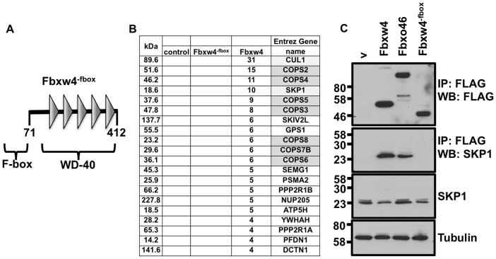
Figure 3. Fbxw4 interacts with E3 ubiquitin ligase components and with the COP9 signalosome in an F-box dependent manner.
A. Schematic of the Fbxw4−fbox protein. Numbers represent the amino acids of the protein. Domains contained within Fbxw4−fbox are indicated. B. Table representing the number of unique peptides identified from one representative mass spectrometry experiment following FLAG immunoprecipitation from lysates of control cells expressing FLAG only “contr.”, or cells expressing FLAG- Fbxw4 or FLAG- Fbxw4−fbox. Column on left indicates the size of the interacting protein, in kilo-daltons (kDa). Gene names of proteins that contain the identified peptides are shown in the right column. Components of an E3 ubiquitin ligase complex are shaded in light gray; components of the COP9 signalosome are shaded dark gray. C. Validation of data mass spectrometry data by immunoprecipitation followed by western blot. 293 T cells were transfected with plasmids containing FLAG-Fbxw4, FLAG-Fbxw4−fbox, FLAG-Fbxo46 or an empty vector (v). 48 hours post-transfection cell lysates were prepared and immunoprecipitations were performed with mono-clonal anti-FLAG antibodies (M2) (to immunoprecipitate Fbxw4-, Fbxw4−fbox-, or Fbxo46-interacting complexes). Western blots were performed to detect Fbxw4, Fbxw4−fbox or Fbxo46 (FLAG rb; polyclonal FLAG antibody; top panel) or SKP1.

