
Fig. 3.
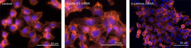
The actin cytoskeleton rearranges after knock-down of cyclin D1 or β-catenin. Immunofluorescences of siRNA treated and phalloidin/TRITC (red) stained HCT116 cells. Control cells were transfected with control siRNA. Nuclei are counterstained with DAPI (blue).