
Fig. 1.
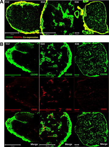
Location of thymic CD248 expressing cells. Frozen sections of E12, 14 and 16 thymuses were co-stained with CD248 (green) and either PDGFRα (A) or CD31 (B) (red). Bar = 100 µm. (For interpretation of the references to colour in this figure legend, the reader is referred to the web version of this article.)