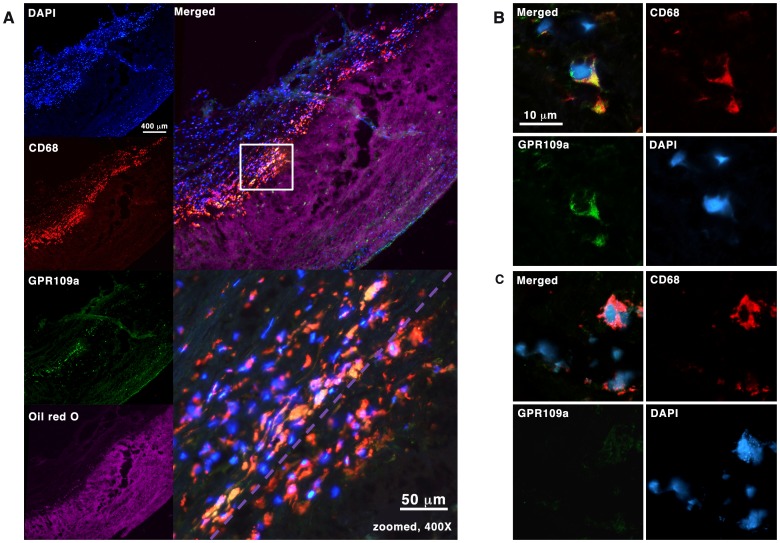
Figure 6. Expression of GPR109A in ex-vivo human carotid plaques.
6A Fluorescence immunohistochemistry staining with antibodies against CD68 and GPR109A. Immediately adjacent 10 µm cryosection was stained with Oil Red O for lipid distribution and visualized using Texas Red excitation filter (540–580 nm) in epifluorescence. CD68-positive cells were seen clustering at the interface between lipid-rich region and the overlying fibrous cap. GPR109A co-expression was seen in a sub-population of these CD68-positive cells outside of the lipid-rich region (yellow). Purple dotted line represents the boundary of the lipid-rich region as seen in the image above. 6B and 6C Loss of GPR109A expression in lipid-rich region. Confocal fluorescence images of CD68-positive cells outside of lipid-rich region are shown in 6B; and those within the lipid-rich region, likely to represent foam cells, are shown in 6C.

