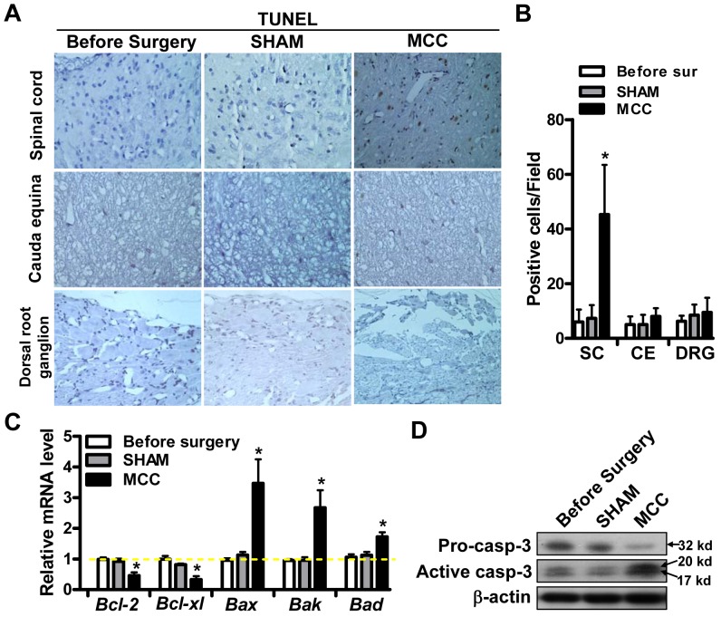
Figure 3. Compression caused apoptosis in spinal cord cells.
(A) The apoptosis of cells in spinal cord (SC), cauda equina (CE) and dorsal root ganglion (DRG) were analyzed by TUNEL method in three groups: before surgery; sham-operated; MCC group. (B) TUNEL positive cells were counted in >5 fields and the average numbers were shown in different groups. (C) qRT-PCR analysis of the expression of Bcl-2, Bcl-xl, Bax, Bak and Bad in three experimental groups. (D) The expression level of pro-caspase-3 (pro-casp-3) and active caspase-3 (active caspase-3) was determined by western blot in indicated 3 groups.

