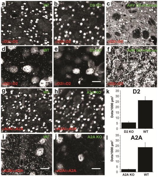
Figure 2. Detection of GPCRs ex vivo in striatal sections using PLA.
D2R was detected in the dorsal striatum of WT mice (a and d). PLA signal from single confocal slice was virtually absent in D2R KO mice (b and e) but was strongly increased when the receptor was overexpressed in the striatum by viral gene transfer of a D2L-R-mVenus (AAV D2-mVenus) (c and f). Similarly A2AR detection in the striatum of WT mice gave a strong PLA signal (g and i) that was absent in A2AR KO mice (h and j). Note that the non-specific nuclear signal (see also SF 3) is similar among genotypes and is unrelated to the presence of primary or secondary antibodies. (k;l) Quantification of PLA signals for D2R (k; unpaired t-test: t= −5.7; p<0.01) and A2AR (l; unpaired t-test: t=−4.2; p<0.01) demonstrates the difference of PLA signal density between WT and KO mice. Scale bars=10 μm.

