Abstract
Background and Aims
Presence of serum antibodies against Mycobacterium avium paratuberculosis (MAP) in Crohn’s Disease (CD) as a disease characteristic remains controversial. In the present work, we assessed antibody reactivity of serum and intestinal fluid against four distinct MAP-antigens, including the recently identified MAP-specific lipopentapeptide (L5P).
Methods
Immunoglobulin concentrations and specificity against 3 non MAP-specific antigens: glycosyl-transferase-d (GSD), purified protein derivative from MAP (Johnin-PPD), heparin binding haemagglutinin (MAP-HBHA) and one MAP-specific antigen: synthetic L5P were determined by ELISA in gut lavage fluids from adult controls or patients with CD, and in sera of children or adult controls or patients with CD, ulcerative colitis or celiac disease.
Results
Total IgA and IgG concentrations were increased in sera of children with CD but were decreased in sera of adults with CD, thereof specificity against MAP antigens was assessed by normalizing immunoglobulin concentrations between samples. In CD patients, IgG reactivity was increased against the four MAP antigens, including L5P in gut lavage fluids but it was only increased against L5P in sera. By contrast, anti-L5P IgG were not increased in patients with ulcerative colitis or celiac disease.
Conclusions
A significant increase in anti-L5P IgG is observed in sera of children and adults with CD but not in patients with other intestinal inflammatory diseases. Anti-L5P antibodies may serve as serological marker for CD.
Introduction
Crohn’s disease (CD) is a multifactorial disease that results from a combination of genetic and environmental risk factors and is associated with exacerbated immune responses against intestinal microbes [1], [2], [3]. The role of specific infectious agents in CD pathogenesis is a long lasting hypothesis. One bacterium that has raised considerable interest is Mycobacterium avium subspecies paratuberculosis (MAP) since Johne’s disease, a chronic enteritis caused by MAP in cattle, shares many clinical and histopathological similarities with CD [4], [5], [6]. As MAP has a very slow growth and is difficult to culture, its putative role in CD has previously been assessed by PCR and serological studies. Despite multiple controversial studies, recent meta-analyses point to a significant association between MAP and CD [7], [8]. MAP belongs to Mycobacterium avium complex (MAC) including M. avium subspecies avium, M. avium subspecies hominissuis and M. avium subspecies silvaticum that are responsible for opportunistic infections in immune-compromised individuals [9], [10], [11], [12]. One important limitation to serological studies has been the absence of a highly specific immunodominant MAP antigen. Surface-located glycopeptidolipids (GPLs), known to participate in the pathogenicity of mycobacteria, have been widely used for serological diagnosis of mycobacterial infections, although they are not strain specific [13], [14], [15]. In MAP, recent genomic and biochemical studies have shown that GPLs are substituted by a specific lipopentapeptide (L5P) [16], [17]. L5P is a major component of the outer part of the cell envelope of MAP and the target of a strong and highly specific IgM and IgG humoral response in MAP-infected animals [16], [17]. So far, L5P has not been tested in serological studies of CD patients. In this study, we have compared systemic and local IgA and IgG antibody responses of children and adults with CD to four different MAP antigens, including synthetic L5P. Due to significant differences in immunoglobulin concentrations between groups of patients, we addressed immunoglobulin specificity by normalizing the immunoglobulin concentration among samples. We observed a significant increase in IgG titers against all four antigens in gut lavage fluids (GLF) of CD patients compared to controls and a specific IgG response against L5P in sera of CD patients compared to controls or to patients with ulcerative colitis or active celiac disease.
Materials and Methods
Patients
In total, 203 sera were analyzed. Ninety two samples were from adults: 45 healthy controls (18–60 years old), 24 CD (mean age: 37 years old, range: 19–78), and 23 active celiac disease (mean age: 38 years old, range: 21–48). 111 samples were from children: 31 healthy controls (mean age: 10 years old, range: 5–16), 47 CD (mean age: 14 years old, range: 8–18), 33 UC (mean age: 11.5 years old, range: 2–17). Sera from adult controls were obtained from the French national blood service (EFS) and sera from pediatric controls were collected during diagnostic workup for a suspected gastro-intestinal disease which was not confirmed. Diagnosis of CD and UC was based on the ECCO and Porto IBD recommendations [18], [19], [20] (combining standard clinical, radiological, endoscopic and histological findings). Diagnosis of active celiac disease was based on the presence of anti-transglutaminase 2 IgA antibodies and on histological criteria (duodenal villous atrophy with increased numbers of intraepithelial lymphocytes) [21].
Gut lavage fluids (GLF) were also collected from 24 adult patients with CD and from 20 control volunteers. Each patient received orally 4 liters of non absorbable polyethylene glycol (PEG)-based lavage fluid (Klean-prep, Norgine). After bowel cleansing was complete, samples of the clear GLF were collected for each patient. Specimens were processed within 30 minutes of collection. Samples were centrifuged (2000 rpm, at 4°C during 5 minutes) and filtered (GF/A, Whatman Scientific Ltd). Soybean trypsin inhibitor (80 µg/ml) (Boerhinger), EDTA (15 mmol/l), phenyl methyl sulfonide fluoride (2 mmol/l), sodium azide (1 mmol/l), and bovine serum albumin were added [22], [23]. Aliquots of GLF were then stored at –70°C.
Local ethics committee approval (CPP Ile de France II, n°C08-31) was obtained for these studies and all patients or their legal guardians signed an informed consent to participate.
Quantification of Total IgA and IgG Concentrations
Sera and GLF IgA and IgG concentrations were measured by ELISA in Immulon 2HB 96-well flat-bottomed plates (Thermo LabSystems) coated overnight at 4°C with 100 µl of 5 µg/ml mouse anti-human IgA or IgG (Abcam) in 0.1 M sodium carbonate coating buffer pH 9.6. Plates were blocked with PBS-5% fetal calf serum (FCS) before incubation with diluted sera (1∶400000 and 1∶106 for IgA and IgG, respectively), diluted GLFs (1∶32000 and 1∶40 for IgA and IgG, respectively), or purified human IgA (MP Bio) or IgG (Biotrend), followed by addition of horseradish-peroxidase (HRP)-conjugated mouse anti-human IgA (Abcam) or IgG (Becton Dickinson) secondary antibodies. HRP was revealed using TMB microwell peroxidase substrate (KPL) and the reaction was stopped with 50 µl 2 N H2SO4 before reading at 450 nm using an automatic Multiskan microplate reader (Thermo LabSystems). Sera and GLF dilutions have been optimized to fit within the range of standard curve; 0.78 to 100 ng/ml for IgA and IgG.
MAP Antigens
Purified protein derivative from MAP (Johnin-PPD) was purchased from the National Veterinary Institute (Oslo, Norway). Synthesized glycosyl transferase d (GSD) epitope 230–244 (15mer TCRRMLAFLKDKENR; mol wt, 1879 Da) was obtained from Covalab. Recombinant heparin binding haemagglutinin (MAP-HBHA) cloned from the genome of the strain MAP K-10 (ATCC BAA-968) was produced in E. coli and purified as previously described [24].
The lipopentapeptide L5P was synthesized manually using the standard Fmoc chemistry protocol on a 4-hydroxymethylbenzoyl resin (HMBA-AM resin, Novabiochem) as described [17].
Immunoglobulin Specificity
Standard antigen titration assays (100 to 1 µg/ml in carbonate buffer for the 4 MAP antigens) were performed to determine the optimal coating concentrations of Johnin-PPD, GSD, L5P or MAP-HBHA. NHS pretreated plates (Covalab) were coated overnight at room temperature with 100 µl of either Johnin-PPD or GSD (20 µg/ml in 0.1 M carbonate buffer). Maxisorp microtiter plates (Nunc, Roskilde, Denmark) were coated overnight at room temperature with 100 µl of L5P (20 µg/ml in Methanol). Immulon 2HB (Thermo LabSystems, Courtaboeuf, France) were coated overnight at 4°C with 100 µl of MAP-HBHA (5 µg/ml in 0.1 M carbonate buffer). After washes with PBS-Tween and blocking in PBS-5% FCS, plates were incubated for 2 hours at 37°C with either a) 100 µl of plasma samples diluted in PBS-5% FCS to adjust IgG concentration to 500 µg/ml for GSD, or 250 µg/ml for L5P and MAP-HBHA, or 125 µg/ml for Johnin-PPD specificity assays or b) 100 µl of GLF samples diluted in PBS-5%FCS to adjust IgG concentration to 250 ng/ml for all of MAP epitopes tested. GLF IgA concentration was adjusted to 18 µg/ml for L5P and MAP-HBHA, 36 µg/ml for Johnin-PPD and 72 µg/ml for GSD. HRP-conjugated mouse anti-human IgG (1∶1000) or IgA (0.325 µg/ml) was incubated for 2 hours at 37°C and revealed as above-mentioned. Results were expressed as OD at 450 nm. Blank wells were loaded with dilution buffer i.e. PBS- 5% FCS. All results were obtained by loading the same amount of immunoglobulin except when results are presented as OD450 nm×dilution factor. In this case, OD450 nm obtained by loading the same amount of IgG are multiplied by the dilution factor required to obtain the appropriate IgG concentration. The OD450 nm multiplied by the dilution factor represents a calculated value of specificity in a same given volume of serum.
Statistical Analysis
Statistical analysis was performed using GraphPad Prism version 5.00 (GraphPad Software, San Diego, California, USA). Results for immunoglobulin concentrations and specificity were compared among groups using the non parametric test Mann-Whitney. Results for IgA and IgG specificity were expressed as OD 450 nm and results were considered positive if above mean optical density (OD) of three blanks (threshold, indicated by a grey dashed line in figures).
Results
IgA and IgG Concentrations in Sera and Gut Lavage Fluids
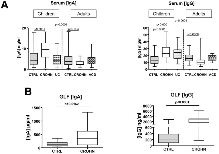
Serum IgA or IgG concentrations were not significantly different between adult and pediatric healthy controls, a result predictable as the mean age of pediatric controls was 10 years [25]. In children, IgA levels were significantly higher in CD patients than in controls whereas IgG levels were significantly higher in either CD or UC patients (Figure 1A). In adults, IgA and IgG levels were significantly lower in CD patients than in controls while no differences were observed between controls and patients with active celiac disease (ACD). As a result, serum IgA and IgG concentrations were significantly higher in children with CD than in adults with CD (3.9 and 2.3 fold increase, respectively).
Figure 1. IgA and IgG concentrations in sera and gut lavage fluids.
(A) Serum IgA and IgG concentrations measured by ELISA in children controls (n = 31), children with CD (n = 47), children with UC (n = 33), adult controls (n = 45), adults with CD (n = 24) and adults with ACD (n = 22). (B) Gut lavage fluids (GLF) IgA and IgG concentrations measured by ELISA in 20 adult controls and 24 adults with CD. The lower and upper lines of the boxes are the 25th and the 75th percentiles, and the lines in the boxes are the median (the 50th percentile). The whiskers extend to the highest and lowest values.
Gut lavage fluids (GLF) from adult CD patients, contained higher IgA and IgG concentrations compared to GLF from controls (2.9 and 8 fold increase, respectively) (Figure 1B). This observation of marked variations in IgA and IgG concentrations between groups of patients might bias comparison of specific serological immune responses between these groups. In order to prevent any misinterpretation of results, each sample has been adjusted according to immunoglobulin concentrations before ELISA.
Increase in Intestinal IgG Against MAP Antigens in Adult CD Patients
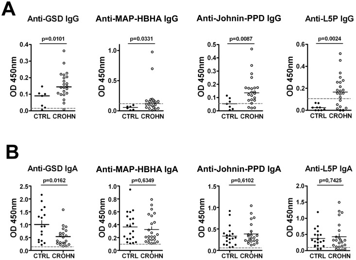
IgG reactivity against all four MAP antigens was significantly higher in GLF from adult CD patients compared to controls (Figure 2A) when normalizing IgG concentration between samples. The difference between CD and controls was particularly striking for L5P as none of the controls showed any signals above threshold while substantial anti-L5P reactivity was observed in 15 out of 22 CD patients. Sensitivity and specificity for anti-L5P IgG in GFL of CD patients were respectively of 70% and 83% (Table S1A). By contrast to IgG, no difference in IgA reactivity against MAP-HBHA, Johnin-PPD or L5P was observed between controls and CD (Figure 2B). Unexpectedly, a significant decrease of anti-GSD IgA was observed in GFL of CD patients compared to controls.
Figure 2. IgG and IgA specificity against MAP-antigens in GLF from adults with or without CD.
(A) IgG specificity assessed by ELISA after normalizing IgG concentration for GSD (7 controls, 22 CD), MAP-HBHA (6 controls, 20 CD), Johnin-PPD (7 controls, 22 CD), L5P (9 controls, 22 CD). (B) IgA specificity assessed by ELISA after normalizing IgA concentration for GSD (16 controls, 20 CD), MAP-HBHA (20 controls, 23 CD), Johnin-PPD (19 controls, 18 CD), L5P (19 controls, 23 CD). Horizontal dashed lines indicate the threshold for specificity corresponding to 3 blanks.
Selective Increase in Serum IgG Against L5P in Children and Adult CD Patients
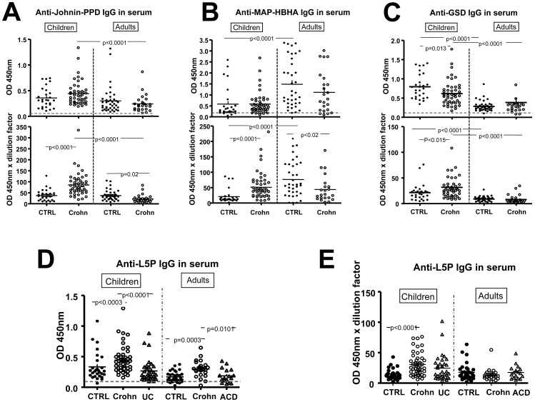
When IgG concentrations were normalized, IgG titers against Johnin-PPD and MAP-HBHA were not significantly different between sera from CD patients and age-matched controls (Figure 3A–B). Unexpectedly, in children only, levels of anti-GSD IgG were significantly lower in CD patients than in controls (Figure 3C). In contrast, anti-L5P IgG titers were significantly higher in both children and adults with CD compared to respective controls (p<0.0003) (Figure 3D). Sensitivity and specificity for anti-L5P IgG are respectively 78% and 59% for adults with CD, and respectively 93% and 48% for children with CD (Table S1-C). In contrast with IgG, no significant increase in IgA levels against L5P, GSD, MAP-HBHA, and Johnin-PPD were detected between CD patients and age-matched controls (Figure S1A–D).
Figure 3. IgG specificity against MAP-antigens in sera of children and adults with or without CD, UC or ACD.
IgG specificity assessed by ELISA after normalizing IgG concentration for (A) Johnin-PPD in children (27 controls, 46 CD) and adults (38 controls, 24 CD). (B) MAP-HBHA in children (28 controls, 47 CD) and adults (38 controls, 23 CD), (C) GSD in children (27 controls, 46 CD) and adults (31 controls, 24 CD), (D) L5P in children (28 controls, 46 CD, 33UC) and adults (32 controls, 23 CD, 17 ACD), (E) Anti-L5P IgG corrected by the dilution factor. Horizontal dashed lines indicate the threshold for specificity corresponding to 3 blanks.
In order to see how variation in serosal IgG concentrations between controls and CD patients (depicted in Figure 1A) could affect the specificity test, we corrected each data by the dilution factor used to obtain normalized IgG concentrations. In children CD patients, IgG specificity against all the tested MAP antigens was then significantly increased (Figure 3A–C). In adult CD patients, specificity against Johnin-PPD and MAP-HBHA was significantly decreased (Figure 3A–B) whereas specificity against GSD and L5P was not significantly different from that of controls (Figure 3C–D). These results are consequences of higher total IgG concentrations in serum of children CD patients, and lower total IgG concentrations in serum of adult CD patients.
In CD patients, anti-L5P IgG titers did not appear to depend on disease location (Table S2) or treatment (Figure S2A–B), but the number of patients may be too small to draw definitive conclusion. However, anti-L5P IgG titers were positively correlated with disease activity based on the Harvey-Bradshaw index in children (p = 0.018), but not with disease activity based on the Crohn’s Disease Activity Index (CDAI) in adults (Figure S3A–B). In both children and adults with CD a positive correlation was also observed between serum IgG titers against L5P and serum IgG titers against Johnin-PPD, GSD or MAP-HBHA respectively (Figure S4). No correlation was observed between total IgG and anti-L5P IgG titers either in children or adults with CD, indicating that these two parameters vary independently (Figure S5A–B).
Importantly, anti-L5P IgG titers were also significantly higher in CD patients than in UC in children (p<0.0001) or ACD in adults (p = 0.0101) (Figure 3D). Sensitivity and specificity of anti-L5P IgG are respectively 78% and 65% for CD adults compared to ACD, and respectively 67% and 36% for CD children compared to UC (Table S1B–C). When anti-L5P IgG titers were corrected with dilution factor, the difference remained only significant between children with CD and pediatric controls (Figure 3E).
Discussion
Taking advantage of the recent description of L5P as a specific MAP antigen that induces strong humoral responses [17], we compared antibody responses to L5P and to three other MAP antigens between Crohn’s disease patients and healthy controls or UC and ACD patients. Antigen specificity is usually measured in the same given volume of fluids (i.e. sera or GLF). However the diversity of immunoglobulin concentrations in GLF and serum might jeopardize the use of a same serum dilution for each sample. The humoral response would be either underestimated in adults with CD, who have fewer antibodies per volume of fluid compared to controls, or overestimated in children with CD, who have more antibodies per volume of fluid compared to control. In this study, the anti-MAP response has been analyzed by normalizing serum immunoglobulin concentrations. IgG responses against all four antigens, were significantly increased in GLF of CD patients, while only anti-L5P IgG were increased in their sera. Since no increased reactivity of plasma IgG against L5P was observed in patients with UC or ACD, using this methodology, anti-L5P IgG may be an interesting serological marker for CD.
Abnormal immune reactivity against microbiota-derived antigens is a characteristic of Crohn’s disease, attested by the detection of serum antibodies against a spectrum of microorganisms. In CD, MAP is one target of the humoral response that raised a lot of interest. A recent meta-analysis concluded to a specific association between the presence of serum anti-MAP antibodies and CD despite wide differences between published studies [7]. The latter differences might result from the use of MAP-sonicates or MAP antigens lacking specificity. To address this issue, we have compared serum reactivity of CD patients against three non-specific MAP antigens and one antigen that has been recently shown to be MAP-specific. The first antigen was Johnin-PPD, a mixture of MAP soluble proteins that has been used for diagnosis of paratuberculosis but showed cross-reactivity with PPD from other mycobacteria [26], [27], [28]. Two other antigens were MAP adhesin HBHA [29] and glycosyl transferase GSD but auto-reactivity of anti-GSD antibodies with intestinal glutathione peroxidase has been suggested [30] as well as cross-reactivity with other mycobacteria [31], [32]. The fourth antigen is a lipopentapeptide (L5P) highly specific for MAP as L5P is only recognized by serum antibodies from animals infected by MAP but not from animals infected by others mycobacteria [17]. Furthermore, in this study ELISA assay was performed with purified synthetic L5P to avoid false positive reactions obtained from native purification preparations likely to be contaminated by non specific MAP cell wall components.
Using GLF to assess intestinal humoral immunity in humans, a 7 fold increase in total IgG but only a 2.5 fold increase in total IgA was observed in GFL from CD compared to controls. In Crohn’s disease, a characteristic of the pathological local humoral response may therefore be the induction of IgG antibodies. Consistent with this hypothesis, previous studies have demonstrated abnormal recruitment of IgG plasma cells in the lamina propria of CD patients [33]. Accumulation of IgG-containing cells has also been observed, in a fewer extent, in gut granulomas of cattle suffering from Johne’s disease [34], [35]. Previous work of A. Fergusson and co-workers demonstrated that IgG in GLF are sensitive marker for CD activity [23], [36]. Indeed, they have reported a significant increase in specific IgG but not IgA against other intraluminal antigens, notably Klebsiella pneumoniae, in gut lavages of CD [36]. In keeping with these results, we observed comparable IgA reactivity against the four antigens in controls and CD patients but significant increase in anti-MAP IgG in CD patients compared to controls. The difference in specific anti-MAP IgG between CD and controls was particularly striking for L5P with 15 of 22 adult CD patients and no adult controls displaying anti-synthetic L5P IgG reactivity above the negative threshold.
The increase in IgG antibodies against all four MAP antigens in CD GLF contrasted with the selective increase in anti-L5P IgG antibodies in CD serum. This increase suggests that increased anti-MAP IgG antibodies in gut lavages are not simply due to plasma leakage but rather result from increased local responses. Specific systemic IgG against microbiota-derived antigens have been ascribed to increased translocation of bacteria or bacteria-derived products in individuals with impaired intestinal barrier [37], [38]. Alternatively, L5P, as compared to others MAP antigens tested, may be more particularly resistant to intestinal and brush border enzymes [16] and/or, as other lipopeptides, possess strong immunogenic properties [39].
Detection of intestinal humoral responses against MAP antigens, and notably against MAP-specific L5P suggests a previous exposure to MAP but is not a direct evidence of a causal role of MAP in CD. Whether MAP can be the triggering infectious agent in a subset of patients remains debated. Recent support to this hypothesis stems from evidence that NOD2 and/or autophagy genes associated with CD participate in the elimination of mycobacteria or in cytokine responses to MAP [40], [41], [42]. Although an association between CARD15/NOD2 polymorphisms and Johne’s disease was reported in cattle [43], no association has been detected between NOD2 mutations and positive MAP serology, in human CD patients. [44]. Thus, it cannot be excluded that intestinal inflammation may promote colonization by MAP, or even that antigens derived from killed-MAP and present in dairy products may induce an IgG response in the inflammatory mucosa of CD patients. Indeed, MAP can be detected in milk of productive livestock. Yet, prevalence of live MAP or MAP-derived antigens in diary food products and human exposure to this bacterium remains a controversial issue [45].
Irrespectively of the exact role of MAP in CD pathogenesis, increased levels of anti-L5P IgG might provide an interesting diagnostic marker for CD. Serum anti-Saccharomyces cerevisiae antibodies (ASCA) and antibodies against other microbiota-derived antigens (OmpC, I2, CBir1) are helpful adjuncts to CD diagnosis. Their specificity has been estimated between 75 to 85% compared to control but their sensitivity is much less and their presence may vary between subsets of patients [46], [47]. Interestingly, ASCA cross react with anti-mycobacteria response [48], [49]. However, ASCA are not specific for CD disease as they can also be detected in ACD [50], [51]. Anti-L5P antibodies may thus represent a valuable specific serological marker for CD as anti-L5P IgG titers were not increased in ACD or UC. Even though, in our present assay, the sensitivity was quite high (78% in adults and 93% in children) the specificity was lower (59% in adults and 48% in children). Yet, the diagnostic value of using L5P for CD (i.e. sensitivy and specificity) might be improved. Indeed, removing the lipid moiety that is not required for L5P recognition [17] or/and coupling L5P to polymers [52], [53] may help to optimize detection of anti-L5P IgG.
In CD children, the increase of total serum IgG might be a consequence of severe systemic inflammation [54]. Accordingly, in this study we observed a positive correlation between anti-L5P IgG titers and clinical score of children (BHI), still those results would need to be confirmed by increasing the number of patients. Moreover, no significant increase of anti-L5P IgG was detected in children with UC despite elevated serum IgG, suggesting that increase of anti-L5P IgG antibodies is specific to CD. However significant differences in anti-L5P IgG titers between CD and UC children patients were only observed when IgG concentrations were normalized. In adults with CD, due to decrease in total serum IgG, a significant increase in serum anti-L5P IgG titers was only observed after adjusting serum IgG concentrations. Those results underline the value of normalizing IgG concentrations when performing an immunoglobulin specificity assay in CD patients.
In conclusion, our work provides evidence of local and systemic IgG response to MAP antigens and notably to the newly defined MAP-specific L5P in Crohn’s disease. Furthermore, our work highlights the value of measuring total immunoglobulin concentration among groups before running antigen specificity assays in order to normalize results and avoid any misinterpretation. Further work is needed to address the diagnostic value of anti-L5P antibodies relative to other serological markers and/or to delineate whether it can define a distinct subgroup of patients with CD.
Supporting Information
IgA specificity against MAP-antigens in sera of children and adults with or without CD. IgA specificity assessed by ELISA after normalizing IgA concentration for (A) L5P in children (26 controls, 42 CD) and adults (31 controls, 22 CD). (B) GSD in children (27 controls, 47 CD) and adults (39 controls, 24 CD), (C) MAP-HBHA in children (28 controls, 46 CD) and adults (35 controls, 24 CD), (D) Johnin-PPD in children (29 controls, 46 CD) and adults (37 controls, 23 CD). Horizontal dashed lines indicate the threshold for specificity corresponding to 3 blanks.
(TIF)
IgG concentrations and IgG specificity against L5P according to treatment of CD patients in children and adults. (A) Serum IgG concentrations measured by ELISA in children and adults with CD. (B) Anti-L5P IgG specificity assessed by ELISA after normalizing IgG concentration in children and adults with CD. n represents the number of patients per treatment. Medications used for treatments were anti-inflammatory drugs (5-ASA), immunosuppressants (Azathioprine, Methotrexate or 6-mercaptopurine) or corticoids (Prednisolone or Budesonide).
(TIF)
Correlations between IgG responses against L5P and disease severity in CD. (A) Correlation between anti-L5P IgG responses obtained after IgG normalization and Harvey-Bradshaw Index (HBI) in children with CD. (B) Correlation between anti-L5P IgG responses obtained after IgG normalization and Crohn’s Disease Activity Index (CDAI) in adults with CD.
(TIF)
Correlations between IgG responses against L5P and other MAP antigens in patients with CD. Spearman’s rank correlation test for correlations between IgG responses against L5P obtained after IgG normalization and other MAP antigens in children (A–C) and adults (D–F) with CD. r : Spearman correlation coefficient.
(TIF)
Correlations between IgG responses against L5P and serum IgG concentrations in patients with CD. Spearman’s rank correlation test for correlations between anti-L5P IgG response obtained after IgG normalization and IgG concentration in children (A) and adults (B).
(TIF)
Sensitivy and specificity of anti-L5P IgG in GLF of adults CD (A), serum of adults with CD (B) and serum of children with CD (C).
(TIF)
Anti-L5P IgG response obtained after IgG normalization (OD450 nm±SEM) according to disease location.
(TIF)
Acknowledgments
The author Jean Marc Reyrat passed away before manuscript submission (October 28, 2009). The rest of the authors acknowledge his contribution to this work. The authors thank Caroline Elie for statistical expertise.
Funding Statement
The authors acknowledge grants from the foundation Princesse Grace de Monaco, the INSERM, Association François Aupetit and Bourses de Recherche Fondamentale sur les Maladies Inflammatoires Chroniques de l’Intestin (BREMICI). The funders had no role in study design, data collection and analysis, decision to publish, or preparation of the manuscript.
References
- 1. Cho JH (2008) The genetics and immunopathogenesis of inflammatory bowel disease. Nat Rev Immunol 8: 458–466. [DOI] [PubMed] [Google Scholar]
- 2. Kaser A, Zeissig S, Blumberg RS (2010) Inflammatory bowel disease. Annu Rev Immunol 28: 573–621. [DOI] [PMC free article] [PubMed] [Google Scholar]
- 3. Nell S, Suerbaum S, Josenhans C (2010) The impact of the microbiota on the pathogenesis of IBD: lessons from mouse infection models. Nat Rev Microbiol 8: 564–577. [DOI] [PubMed] [Google Scholar]
- 4. Chiodini RJ (1989) Crohn’s disease and the mycobacterioses: a review and comparison of two disease entities. Clin Microbiol Rev 2: 90–117. [DOI] [PMC free article] [PubMed] [Google Scholar]
- 5. Greenstein RJ (2003) Is Crohn’s disease caused by a mycobacterium? Comparisons with leprosy, tuberculosis, and Johne’s disease. Lancet Infect Dis 3: 507–514. [DOI] [PubMed] [Google Scholar]
- 6. Behr MA, Kapur V (2008) The evidence for Mycobacterium paratuberculosis in Crohn’s disease. Curr Opin Gastroenterol 24: 17–21. [DOI] [PubMed] [Google Scholar]
- 7. Feller M, Huwiler K, Stephan R, Altpeter E, Shang A, et al. (2007) Mycobacterium avium subspecies paratuberculosis and Crohn’s disease: a systematic review and meta-analysis. Lancet Infect Dis 7: 607–613. [DOI] [PubMed] [Google Scholar]
- 8. Chassaing B, Darfeuille-Michaud A (2011) The commensal microbiota and enteropathogens in the pathogenesis of inflammatory bowel diseases. Gastroenterology 140: 1720–1728. [DOI] [PubMed] [Google Scholar]
- 9. Thorel MF, Krichevsky M, Levy-Frebault VV (1990) Numerical taxonomy of mycobactin-dependent mycobacteria, emended description of Mycobacterium avium, and description of Mycobacterium avium subsp. avium subsp. nov., Mycobacterium avium subsp. paratuberculosis subsp. nov., and Mycobacterium avium subsp. silvaticum subsp. nov. Int J Syst Bacteriol 40: 254–260. [DOI] [PubMed] [Google Scholar]
- 10. Mijs W, de Haas P, Rossau R, Van der Laan T, Rigouts L, et al. (2002) Molecular evidence to support a proposal to reserve the designation Mycobacterium avium subsp. avium for bird-type isolates and ’M. avium subsp. hominissuis’ for the human/porcine type of M. avium. Int J Syst Evol Microbiol 52: 1505–1518. [DOI] [PubMed] [Google Scholar]
- 11. Turenne CY, Wallace R Jr, Behr MA (2007) Mycobacterium avium in the postgenomic era. Clin Microbiol Rev 20: 205–229. [DOI] [PMC free article] [PubMed] [Google Scholar]
- 12. Primm TP, Lucero CA, Falkinham JO 3rd (2004) Health impacts of environmental mycobacteria. Clin Microbiol Rev 17: 98–106. [DOI] [PMC free article] [PubMed] [Google Scholar]
- 13. Nishimura T, Hasegawa N, Fujita Y, Yano I, Ishizaka A (2009) Serodiagnostic contributions of antibody titers against mycobacterial lipid antigens in Mycobacterium avium complex pulmonary disease. Clin Infect Dis 49: 529–535. [DOI] [PubMed] [Google Scholar]
- 14. Kitada S, Maekura R, Toyoshima N, Fujiwara N, Yano I, et al. (2002) Serodiagnosis of pulmonary disease due to Mycobacterium avium complex with an enzyme immunoassay that uses a mixture of glycopeptidolipid antigens. Clin Infect Dis 35: 1328–1335. [DOI] [PubMed] [Google Scholar]
- 15. Fujita Y, Doi T, Maekura R, Ito M, Yano I (2006) Differences in serological responses to specific glycopeptidolipid-core and common lipid antigens in patients with pulmonary disease due to Mycobacterium tuberculosis and Mycobacterium avium complex. J Med Microbiol 55: 189–199. [DOI] [PubMed] [Google Scholar]
- 16. Eckstein TM, Chandrasekaran S, Mahapatra S, McNeil MR, Chatterjee D, et al. (2006) A major cell wall lipopeptide of Mycobacterium avium subspecies paratuberculosis. J Biol Chem 281: 5209–5215. [DOI] [PubMed] [Google Scholar]
- 17. Biet F, Bay S, Thibault VC, Euphrasie D, Grayon M, et al. (2008) Lipopentapeptide induces a strong host humoral response and distinguishes Mycobacterium avium subsp. paratuberculosis from M. avium subsp. avium. Vaccine 26: 257–268. [DOI] [PubMed] [Google Scholar]
- 18. Inflammatory bowel disease in children and adolescents: recommendations for diagnosis–the Porto criteria. J Pediatr Gastroenterol Nutr 41: 1–7. [DOI] [PubMed] [Google Scholar]
- 19. Travis SP, Stange EF, Lemann M, Oresland T, Bemelman WA, et al. (2008) European evidence-based Consensus on the management of ulcerative colitis: Current management. J Crohns Colitis 2: 24–62. [DOI] [PubMed] [Google Scholar]
- 20. Van Assche G, Dignass A, Panes J, Beaugerie L, Karagiannis J, et al. (2010) The second European evidence-based Consensus on the diagnosis and management of Crohn’s disease: Definitions and diagnosis. J Crohns Colitis 4: 7–27. [DOI] [PubMed] [Google Scholar]
- 21. Revised criteria for diagnosis of coeliac disease. Report of Working Group of European Society of Paediatric Gastroenterology and Nutrition. Arch Dis Child 65: 909–911. [DOI] [PMC free article] [PubMed] [Google Scholar]
- 22. Gaspari MM, Brennan PT, Solomon SM, Elson CO (1988) A method of obtaining, processing, and analyzing human intestinal secretions for antibody content. J Immunol Methods 110: 85–91. [DOI] [PubMed] [Google Scholar]
- 23. O’Mahony S, Barton JR, Crichton S, Ferguson A (1990) Appraisal of gut lavage in the study of intestinal humoral immunity. Gut 31: 1341–1344. [DOI] [PMC free article] [PubMed] [Google Scholar]
- 24. Pethe K, Aumercier M, Fort E, Gatot C, Locht C, et al. (2000) Characterization of the heparin-binding site of the mycobacterial heparin-binding hemagglutinin adhesin. J Biol Chem 275: 14273–14280. [DOI] [PubMed] [Google Scholar]
- 25. Manz RA, Hauser AE, Hiepe F, Radbruch A (2005) Maintenance of serum antibody levels. Annu Rev Immunol 23: 367–386. [DOI] [PubMed] [Google Scholar]
- 26. Konst H, McIntosh CW (1958) Studies Of Johne’s Disease In Canada. I. The Use Of Johnin P.P.D. Can J Comp Med Vet Sci 22: 157–160. [PMC free article] [PubMed] [Google Scholar]
- 27. Koets AP, Rutten VP, de Boer M, Bakker D, Valentin-Weigand P, et al. (2001) Differential changes in heat shock protein-, lipoarabinomannan-, and purified protein derivative-specific immunoglobulin G1 and G2 isotype responses during bovine Mycobacterium avium subsp. paratuberculosis infection. Infect Immun 69: 1492–1498. [DOI] [PMC free article] [PubMed] [Google Scholar]
- 28. Benjamin RG, Debanne SM, Ma Y, Daniel TM (1984) Evaluation of mycobacterial antigens in an enzyme-linked immunosorbent assay (ELISA) for the serodiagnosis of tuberculosis. J Med Microbiol 18: 309–318. [DOI] [PubMed] [Google Scholar]
- 29. Menozzi FD, Rouse JH, Alavi M, Laude-Sharp M, Muller J, et al. (1996) Identification of a heparin-binding hemagglutinin present in mycobacteria. J Exp Med 184: 993–1001. [DOI] [PMC free article] [PubMed] [Google Scholar]
- 30. Polymeros D, Bogdanos DP, Day R, Arioli D, Vergani D, et al. (2006) Does cross-reactivity between mycobacterium avium paratuberculosis and human intestinal antigens characterize Crohn’s disease? Gastroenterology 131: 85–96. [DOI] [PubMed] [Google Scholar]
- 31. Tizard M, Bull T, Millar D, Doran T, Martin H, et al. (1998) A low G+C content genetic island in Mycobacterium avium subsp. paratuberculosis and M. avium subsp. silvaticum with homologous genes in Mycobacterium tuberculosis. Microbiology 144 (Pt 12): 3413–3423. [DOI] [PubMed] [Google Scholar]
- 32. Sechi LA, Ahmed N, Felis GE, Dupre I, Cannas S, et al. (2006) Immunogenicity and cytoadherence of recombinant heparin binding haemagglutinin (HBHA) of Mycobacterium avium subsp. paratuberculosis: functional promiscuity or a role in virulence? Vaccine 24: 236–243. [DOI] [PubMed] [Google Scholar]
- 33. Brandtzaeg P, Halstensen TS, Kett K, Krajci P, Kvale D, et al. (1989) Immunobiology and immunopathology of human gut mucosa: humoral immunity and intraepithelial lymphocytes. Gastroenterology 97: 1562–1584. [DOI] [PubMed] [Google Scholar]
- 34. Momotani E, Ishikawa Y, Yoshino T (1986) Immunohistochemical distribution of immunoglobulin and secretory component in the ileum of normal and paratuberculosis-infected cattle. J Comp Pathol 96: 659–669. [DOI] [PubMed] [Google Scholar]
- 35. Momotani E, Romona NM, Yoshihara K, Momotani Y, Hori M, et al. (2012) Molecular pathogenesis of bovine paratuberculosis and human inflammatory bowel diseases. Vet Immunol Immunopathol 148: 55–68. [DOI] [PubMed] [Google Scholar]
- 36. O’Mahony S, Anderson N, Nuki G, Ferguson A (1992) Systemic and mucosal antibodies to Klebsiella in patients with ankylosing spondylitis and Crohn’s disease. Ann Rheum Dis 51: 1296–1300. [DOI] [PMC free article] [PubMed] [Google Scholar]
- 37. Macpherson AJ, Uhr T (2004) Induction of protective IgA by intestinal dendritic cells carrying commensal bacteria. Science 303: 1662–1665. [DOI] [PubMed] [Google Scholar]
- 38. Konrad A, Cong Y, Duck W, Borlaza R, Elson CO (2006) Tight mucosal compartmentation of the murine immune response to antigens of the enteric microbiota. Gastroenterology 130: 2050–2059. [DOI] [PubMed] [Google Scholar]
- 39. Abdel-Aal AB, Batzloff MR, Fujita Y, Barozzi N, Faria A, et al. (2008) Structure-activity relationship of a series of synthetic lipopeptide self-adjuvanting group a streptococcal vaccine candidates. J Med Chem 51: 167–172. [DOI] [PubMed] [Google Scholar]
- 40. Ferwerda G, Girardin SE, Kullberg BJ, Le Bourhis L, de Jong DJ, et al. (2005) NOD2 and toll-like receptors are nonredundant recognition systems of Mycobacterium tuberculosis. PLoS Pathog 1: 279–285. [DOI] [PMC free article] [PubMed] [Google Scholar]
- 41. Ferwerda G, Kullberg BJ, de Jong DJ, Girardin SE, Langenberg DM, et al. (2007) Mycobacterium paratuberculosis is recognized by Toll-like receptors and NOD2. J Leukoc Biol 82: 1011–1018. [DOI] [PubMed] [Google Scholar]
- 42. Jostins L, Ripke S, Weersma RK, Duerr RH, McGovern DP, et al. (2012) Host-microbe interactions have shaped the genetic architecture of inflammatory bowel disease. Nature 491: 119–124. [DOI] [PMC free article] [PubMed] [Google Scholar]
- 43. Pinedo PJ, Buergelt CD, Donovan GA, Melendez P, Morel L, et al. (2009) Association between CARD15/NOD2 gene polymorphisms and paratuberculosis infection in cattle. Vet Microbiol 134: 346–352. [DOI] [PubMed] [Google Scholar]
- 44. Bernstein CN, Wang MH, Sargent M, Brant SR, Collins MT (2007) Testing the interaction between NOD-2 status and serological response to Mycobacterium paratuberculosis in cases of inflammatory bowel disease. J Clin Microbiol 45: 968–971. [DOI] [PMC free article] [PubMed] [Google Scholar]
- 45. Gill CO, Saucier L, Meadus WJ (2011) Mycobacterium avium subsp. paratuberculosis in dairy products, meat, and drinking water. J Food Prot 74: 480–499. [DOI] [PubMed] [Google Scholar]
- 46. Peyrin-Biroulet L, Standaert-Vitse A, Branche J, Chamaillard M (2007) IBD serological panels: facts and perspectives. Inflamm Bowel Dis 13: 1561–1566. [DOI] [PubMed] [Google Scholar]
- 47. Ruemmele FM, Targan SR, Levy G, Dubinsky M, Braun J, et al. (1998) Diagnostic accuracy of serological assays in pediatric inflammatory bowel disease. Gastroenterology 115: 822–829. [DOI] [PubMed] [Google Scholar]
- 48. Cohavy O, Harth G, Horwitz M, Eggena M, Landers C, et al. (1999) Identification of a novel mycobacterial histone H1 homologue (HupB) as an antigenic target of pANCA monoclonal antibody and serum immunoglobulin A from patients with Crohn’s disease. Infect Immun 67: 6510–6517. [DOI] [PMC free article] [PubMed] [Google Scholar]
- 49. Muller S, Schaffer T, Schoepfer AM, Hilty A, Bodmer T, et al. (2008) Partial overlap of anti-mycobacterial, and anti-Saccharomyces cerevisiae mannan antibodies in Crohn’s disease. World J Gastroenterol 14: 3650–3661. [DOI] [PMC free article] [PubMed] [Google Scholar]
- 50. Kotze LM, Nisihara RM, Utiyama SR, Kotze PG, Theiss PM, et al. (2010) Antibodies anti-Saccharomyces cerevisiae (ASCA) do not differentiate Crohn’s disease from celiac disease. Arq Gastroenterol 47: 242–245. [DOI] [PubMed] [Google Scholar]
- 51. Biet F, Gendt L, Anton E, Ballot E, Hugot JP, et al. (2011) Serum Antibodies to Mycobacterium avium Subspecies paratuberculosis Combined with Anti-Saccharomyces cerevisiae Antibodies in Crohn’s Disease Patients: Prevalence and Diagnostic Role. Dig Dis Sci 56: 1794–1800. [DOI] [PubMed] [Google Scholar]
- 52. Cartwright GA, Rothel JS, Lightowlers MW (1995) Conventional immunoassays underestimate anti-GST antibody titre. J Immunol Methods 179: 31–35. [DOI] [PubMed] [Google Scholar]
- 53. Qiu B, Brunner M, Zhang G, Sigal L, Stein S (2000) Selection of continuous epitope sequences and their incorporation into poly(ethylene glycol)-peptide conjugates for use in serodiagnostic immunoassays: application to Lyme disease. Biopolymers 55: 319–333. [DOI] [PubMed] [Google Scholar]
- 54. Ruemmele FM (2010) Pediatric inflammatory bowel diseases: coming of age. Curr Opin Gastroenterol 26: 332–336. [DOI] [PubMed] [Google Scholar]
Associated Data
This section collects any data citations, data availability statements, or supplementary materials included in this article.
Supplementary Materials
IgA specificity against MAP-antigens in sera of children and adults with or without CD. IgA specificity assessed by ELISA after normalizing IgA concentration for (A) L5P in children (26 controls, 42 CD) and adults (31 controls, 22 CD). (B) GSD in children (27 controls, 47 CD) and adults (39 controls, 24 CD), (C) MAP-HBHA in children (28 controls, 46 CD) and adults (35 controls, 24 CD), (D) Johnin-PPD in children (29 controls, 46 CD) and adults (37 controls, 23 CD). Horizontal dashed lines indicate the threshold for specificity corresponding to 3 blanks.
(TIF)
IgG concentrations and IgG specificity against L5P according to treatment of CD patients in children and adults. (A) Serum IgG concentrations measured by ELISA in children and adults with CD. (B) Anti-L5P IgG specificity assessed by ELISA after normalizing IgG concentration in children and adults with CD. n represents the number of patients per treatment. Medications used for treatments were anti-inflammatory drugs (5-ASA), immunosuppressants (Azathioprine, Methotrexate or 6-mercaptopurine) or corticoids (Prednisolone or Budesonide).
(TIF)
Correlations between IgG responses against L5P and disease severity in CD. (A) Correlation between anti-L5P IgG responses obtained after IgG normalization and Harvey-Bradshaw Index (HBI) in children with CD. (B) Correlation between anti-L5P IgG responses obtained after IgG normalization and Crohn’s Disease Activity Index (CDAI) in adults with CD.
(TIF)
Correlations between IgG responses against L5P and other MAP antigens in patients with CD. Spearman’s rank correlation test for correlations between IgG responses against L5P obtained after IgG normalization and other MAP antigens in children (A–C) and adults (D–F) with CD. r : Spearman correlation coefficient.
(TIF)
Correlations between IgG responses against L5P and serum IgG concentrations in patients with CD. Spearman’s rank correlation test for correlations between anti-L5P IgG response obtained after IgG normalization and IgG concentration in children (A) and adults (B).
(TIF)
Sensitivy and specificity of anti-L5P IgG in GLF of adults CD (A), serum of adults with CD (B) and serum of children with CD (C).
(TIF)
Anti-L5P IgG response obtained after IgG normalization (OD450 nm±SEM) according to disease location.
(TIF)


