
Figure 4. Isolation of iNOS binding partners unique to the different subcellular populations.
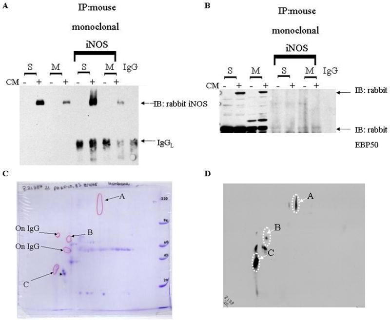
The different treatment groups were separated into subcellular fractions and immunoprecipitated (IP) (METHODS). A. Immunoprecipitation reactions with monoclonal iNOS was WB positive for EBP50. B. Immunoprecipitation reactions with monoclonal EBP50 was iNOS immunonegative. Immunoprecipitation reactions of the Soluble, Membrane and IgG samples were separated on an 8% 2-D gel (Kendrick Laboratory). C. Coomassie stained gel compared to a duplicated gel that was probed for iNOS. D. Spot B is the only iNOS positive spot unique to the membrane IP 2-D gel. This sample was excised, trypsin digested, analyzed by MALDI-MS (M. Gawinowicz, Columbia University) and identified as actin. Spots A and C are found in both 2-D gels and C has been identified as EBP50. The IgG 2-D gel was also probed for iNOS and positive spots were excluded from further characterization. (−)= media; (+)= cytokine mixture (IL-1, RGI, TNFα); S=supernatant of 100,000xg; M=pellet of 100,000xg.