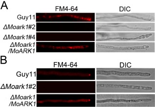
Figure 1.

MoArk1 has a role in the formation of the Spitzenkörper and endocytosis. (A) FM4‐64 staining revealed that the ΔMoark1 mutant was defective in endocytosis. Strains were grown for 2 days on complete medium (CM)‐overlaid microscope slides before the addition of FM4‐64 and photographs were taken after 15 min of exposure to FM4‐64. Camera exposure is indicated in seconds (800 ms). (B) The wild‐type and complemented strains showed the presence of an intact Spitzenkörper at the tips of the hyphae, which was missing in the ΔMoark1 mutants after exposure to FM4‐64 staining for 15 min. Strains were grown for 2 days on CM‐overlaid microscope slides before staining. DIC, differential interference contrast image.
