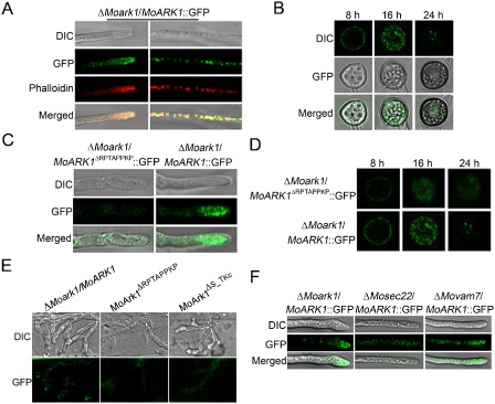
Figure 7.

Cellular localization of MoArk1‐green fluorescent protein (MoArk1‐GFP) and MoArk1 Δ RPTAPPKP‐GFP. (A) Vegetative hyphal tips were observed under laser scanning confocal microscopy. The hyphae were stained with the actin dye rhodamine‐phalloidin after 48 h of static culture. DIC, differential interference contrast image. (B) ΔMoark1/ MoArk1‐GFP fluorescence observation during appressoria formation process after 8, 16 and 24 h under laser scanning confocal microscopy. (C) Vegetative hyphal tips of ΔMoark1/MoArk1 Δ RPTAPPKP‐GFP were observed under laser scanning confocal microscopy. (D) ΔMoark1/MoArk1 Δ RPTAPPKP‐GFP fluorescence observation during appressoria formation process after 8, 16 and 24 h under laser scanning confocal microscopy. (E) Invasive hyphae produced by the MoARK1‐GFP, MoARK1 Δ RPTAPPKP‐GFP and MoARK1 Δ S _ TKc‐GFP transformants in onion epidermal cells at 24 hpi. (F) Vegetative hyphal tips of ΔMoark1/MoARK1, ΔMosec22/MoARK1 and ΔMovam7/MoARK1 under laser scanning confocal microscopy.
