
Figure 1.
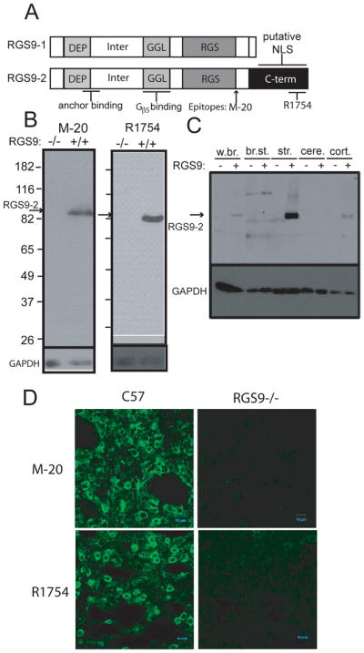
A. Domain structures of the RGS9-1 and RGS9-2 proteins and location of RGS9-2 specific antibody (M-20 and R1754) epitopes. DEP= disheveled, egl-10, plekstrin homology; Inter=area between the DEP and GGL domains; GGL= g gamma-like; RGS= regulator of G-protein signaling; C-term = RGS9-2 c-terminus; NLS = nuclear localization signal. B. Testing the specificity of anti-RGS9-2 antibodies by immunoblot. Both the M-20 and R1754 antibodies recognize a single band of approximately 82 kD in mouse striatal homogenate that is absent in the RGS9 −/− mouse. C. Immunoblot for RGS9-2 in a various brain tissue homogenates shows that RGS9-2 is present in the brain and highly enriched in the striatum. w.br. = whole brain; br.st. = brain stem; str. = striatum; cere. = cerebellum; cort. = cortex. D. Immunoflurescence staining of mouse striatal slices from C57/Bl6 and RGS9−/− using both RGS9-2 specific antibodies (M-20 and R1754). All signal is specific to RGS9-2 as it is absent in RGS9−/−.