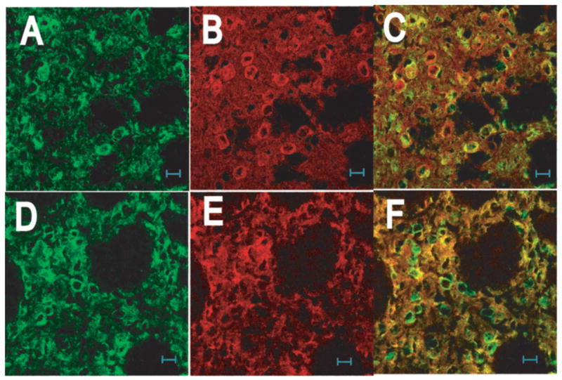
Figure 2.

Cellular and subcellular distribution of RGS9-2 in striatum. Sections from wildtype mouse were sequentially stained with either the goat anti-RGS9-2 M20 and mouse anti-DARPP32 antibody (A –C) or the goat anti-RGS9-2 antibody and rabbit anti-spinophilin antibody (D –F). No heat-induced antigen retrieval treatment was used as such treatment abolished the staining for DARPP32. Images in the left (green), middle (red), and right columns are for RGS9-2, marker, and merges immunofluorescence, respectively.
