Figure 5.
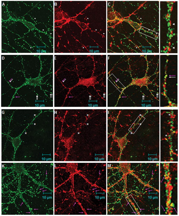
RGS9-2 immunofluorescence is found postsynaptically along the dendritic shaft in cultured striatal neurons. Rat striatal-cortical co-cultures were double stained for RGS9-2 and synaptophysin (A–C, 14DIV), or PSD-95 (D–F, 14DIV), or spinophilin (G–I, 14DIV and J–L, 21DIV). Images in the left (green), middle (red), and right columns are for RGS9-2, marker, and merged immunofluorescence, respectively. In A–C, axons outlined by synaptophysin-positive varicosities intertwine with dendrites outlined by the RGS9-2 positive puncta. RGS9-2 immunostaining are found opposed the synaptophysin immunoreactive puncta (arrowheads). In D–F, the discrete staining pattern of RGS9-2 immunoreactivity and that of PSD95 overlaps to a great extent. White arrows indicate areas of co-localization. The pink arrows indicate RGS9-2/PSD95 puncta that exhibit a spine like structure. In G–I, RGS9-2 immunostaining does not share the same pattern with spinophilin. Note there are a few puncta (marked by asterisks) of RGS9-2 found at the base of the dendritic protrusions. In K-L, spinophilin antibody labels the mature form of spine in 21DIV co-cultures. Very few RGS9-2 positive puncta co-localize with spinophilin positive spines (pink arrows). The majority of RGS9-2 positive puncta remained positioned along the dendritic shaft. Boxed areas are a 2.67x enlargement.

