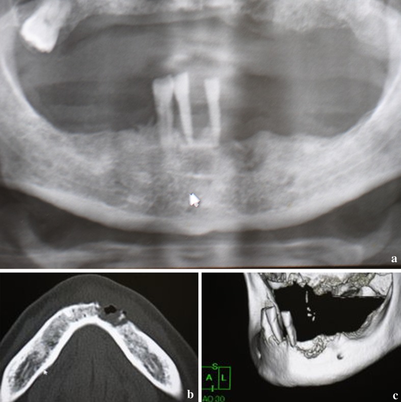
Fig. 2.

Radiologic findings before therapy. a Panoramic radiograph showing irregular radiolucent lesion in the mandible located around the site of canine extraction. b CT showing marked cortical bone osteolysis with sequestrated loose bony tissue. c 3D-CT view showing a so-called “moth-eaten” irregular bone resorptive surface, and remaining teeth with severe periodontitis
