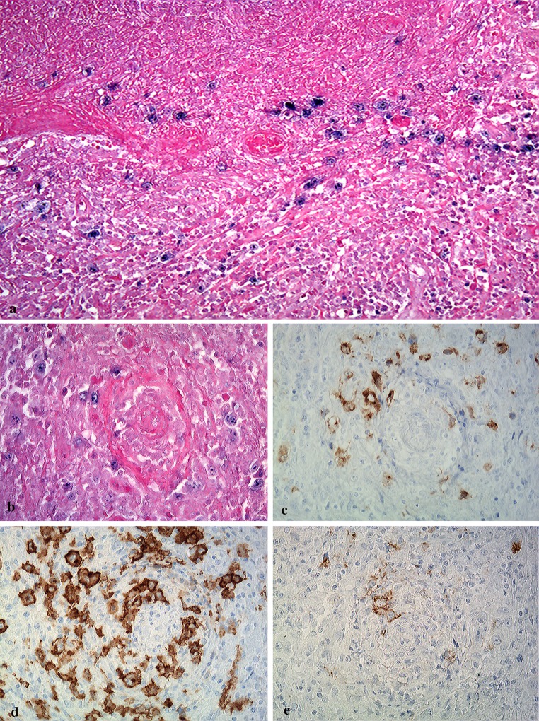
Fig. 6.
Distribution of EBV + large atypical lymphoid cells in hemorrhagic necrosis and granulomatous areas. EBER-positive cells showing angiocentric/angiodestructive pattern are present in areas of hemorrhagic necrosis (a) and granulomatous formation (b) (original magnification ×100, and ×200). Large atypical lymphoid cells in perivascular or vascular areas are positive for LMP-1 (c), CD30 (d), and CD20 (e) (original magnification ×200)

