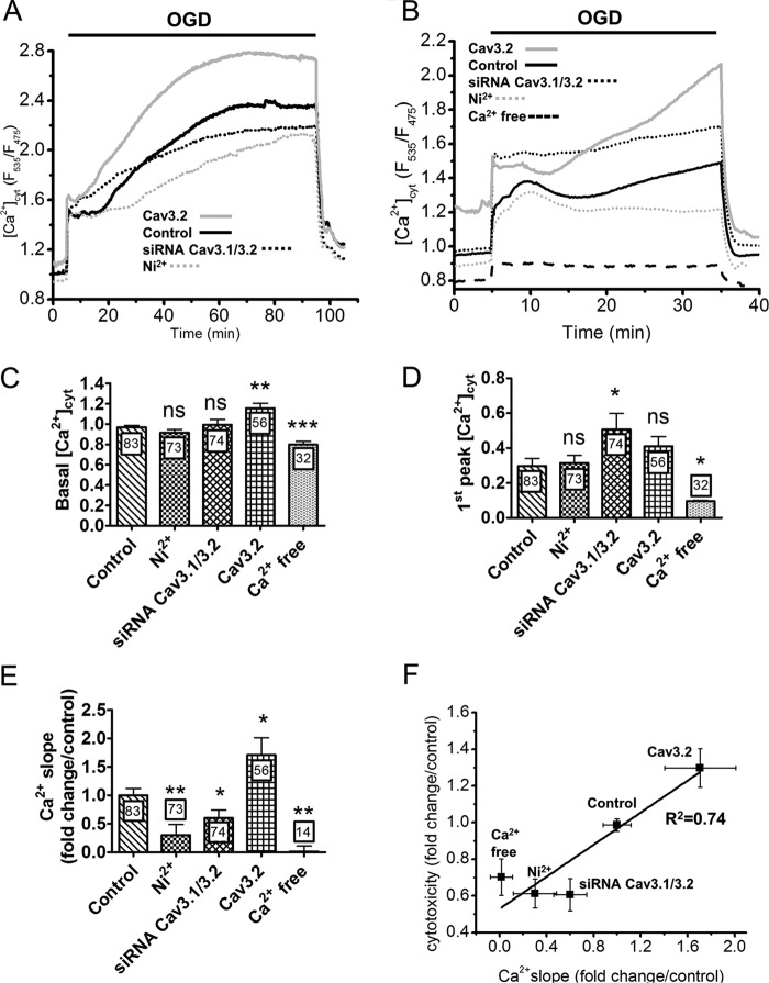
FIGURE 3.
Effect of T-type Ca2+ channels modulation on the [Ca2+]cyt elevations evoked by OGD. A, [Ca2+]cyt recordings during 90 min of OGD. The delayed [Ca2+]cyt elevation was reduced by Ni2+ and Cav3.1/Cav3.2 silencing and increased by Cav3.2 overexpression. Traces are the averaged [Ca2+]cyt responses measured in the indicated conditions. B, [Ca2+]cyt elevations evoked by 30 min of OGD. External Ca2+ removal prevented the elevations. C, basal [Ca2+]cyt levels recorded under the different conditions. D, amplitude of the initial [Ca2+]cyt elevation evoked by OGD. Data are mean ± S.E. from six to eight independent recordings. The total number of measured cells is indicated inside the bars. E, effect of T-type Ca2+ channel modulation on the slope of the secondary [Ca2+]cyt elevation. Data are mean ± S.E. from three to four independent recordings, expressed as fold changes from the control condition. The total number of measured cells is indicated inside the bars. F, cytotoxicity as a function of the Ca2+ slope measured in the indicated conditions. Data are from Figs. 2A and 3E. ns, non-significant; *, p < 0.05; **, p < 0.01; ***, p < 0.001 (Student's t test for unpaired samples).

