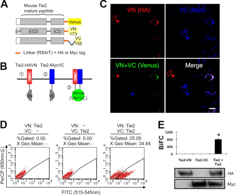
FIGURE 1.
BiFC analysis of Tie2 receptor homodimerization in living cells. A and B, schematic representation of Tie2 tagged with either the N- or C-terminal of the Venus fragment (VN or VC). SP, signal peptide; ECD, extracellular domain; ICD, intracellular domain. When Tie2 dimerizes, fluorescence should reconstitute. C, HEK293T cells expressing Tie2-HAVN (red) and Tie2-MycVC (blue) observed by confocal microscopy. Cells were co-transefected with Tie2-HAVN and Tie2-MycVC expression vectors. Note that cells expressing Tie2-HAVN alone (red arrowhead) or Tie2-MycVC alone (blue arrowhead) develop no Venus fluorescence. Bar indicates 20 μm. D, flow cytometric analysis for evaluation of receptor dimerization as indicated. E, quantitative evaluation of Tie2 homodimerization in BiFC as observed in D (*, p < 0.05; n = 3). Protein expression level of each receptor was assessed by immunoblotting with anti-HA or anti-Myc Ab.

