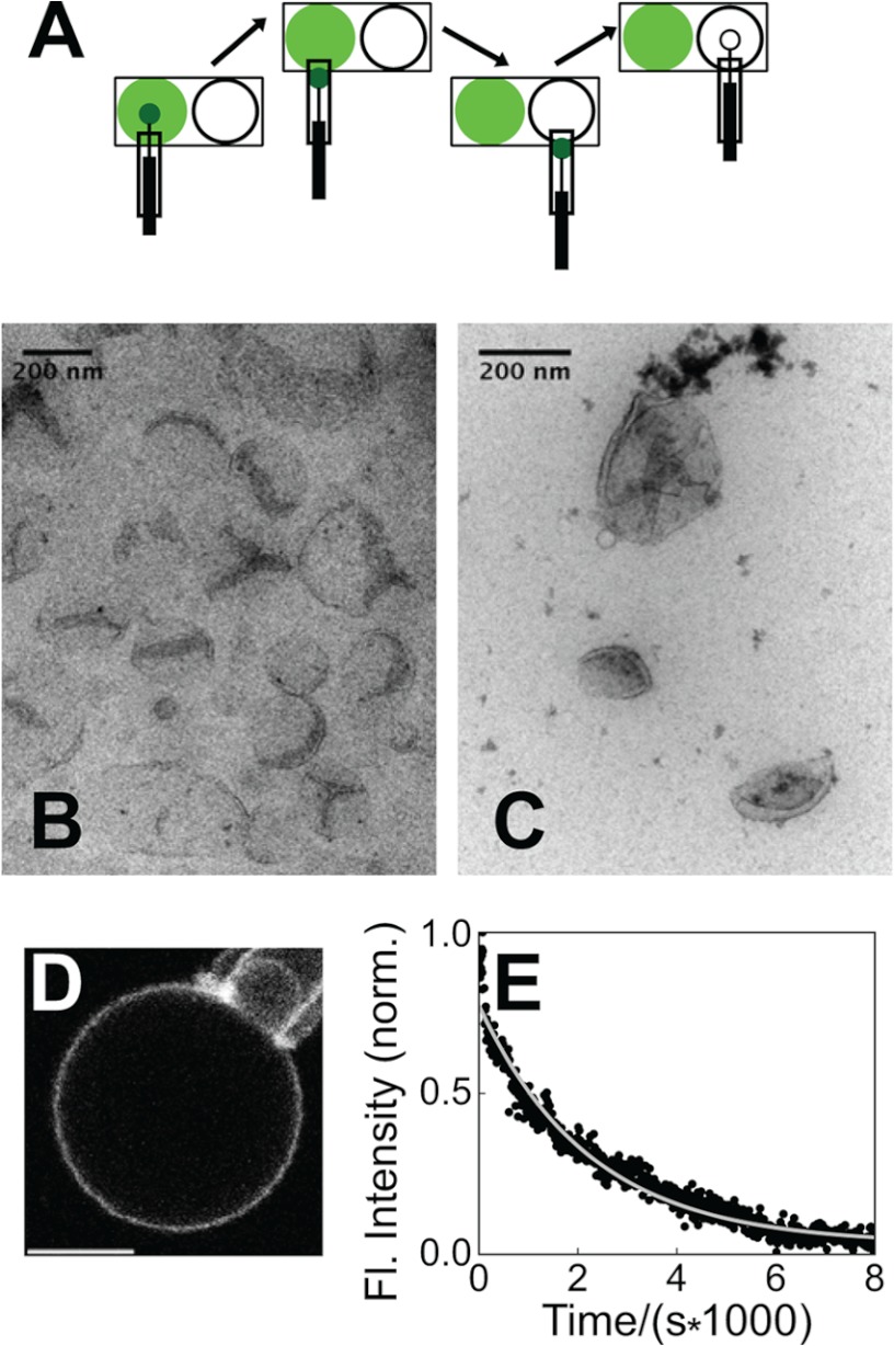
FIGURE 4.
Dissociation kinetics under nontubulating conditions. A, diagram of dissociation kinetics experiments employing rapid dilution via micropipette-mediated GUV transfer. B and C, electron micrographs of liposomes (composed of 25% DOPG) in the absence of endophilin N-BAR (B) and in the presence of 300 nm N-BAR_C241-AF488 (4 μm lipids, 33 mm NaCl) (C). D, fluorescence micrograph of N-BAR_241-AF-488 bound to a GUV (25% DOPG) aspirated in a micropipette, in 33 mm NaCl, 1 mm DTT following preincubation at 300 nm protein, 4 μm lipids, and identical ionic strength. Scale bar, 5 μm. E, fluorescence (Fl.) intensity record and single-exponential fit (solid line) charting dissociation of N-BAR, as diagrammed in A, with conditions of D. norm., normalized.

