FIGURE 8.
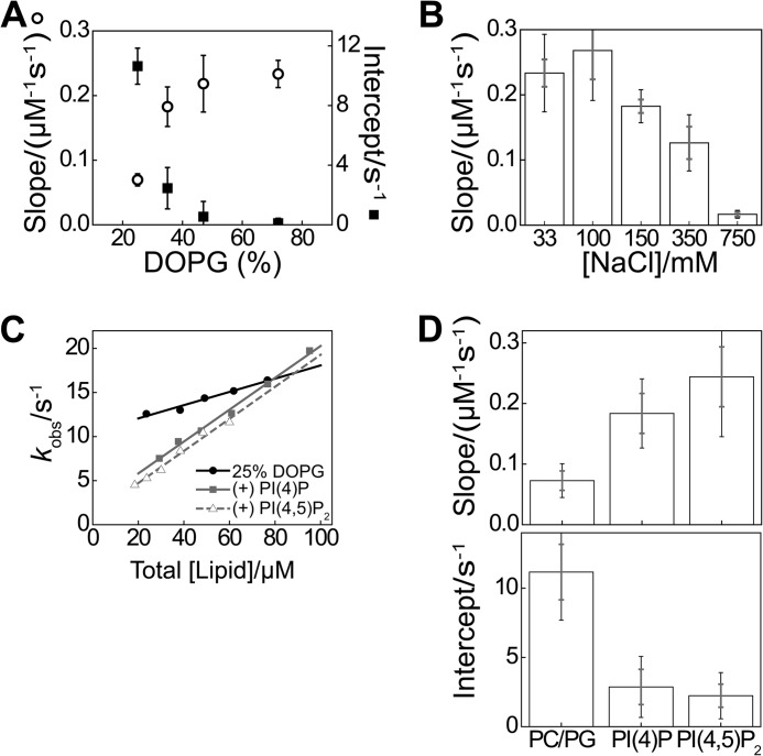
Prominent role of electrostatics in the first step of the membrane binding mechanism. A, parameters extracted from analysis of titrations as shown in Fig. 5C, for indicated membrane compositions, averaged over 3–7 vesicle preparations (error bars indicate S.E.), with 33 mm NaCl and 0.3 μm N-BAR A66W. B, averaged slope as in A for experiments varying solution ionic strength via the NaCl concentration, with 72% DOPG and 0.3 μm N-BAR A66W (error bars indicate S.D. and S.E. over ≥3 vesicle preparations). C, DPH-PC stopped-flow association analysis in the pseudo-first-order regime, using 0.3 μm N-BAR A66W, 33 mm NaCl, with 25% DOPG and phosphoinositides (PI(4)P and PI(4,5)P2) incorporated to 3 mol %. D, pseudo-first-order parameters extracted from plots exemplified in C. Error bars represent S.D. and S.E. of datasets as in C from ≥3 vesicle preparations. A similar trend was observed using 150 mm NaCl and a background composition of 35% DOPG (data not shown).

