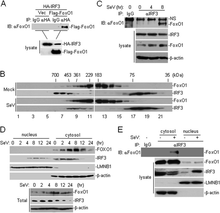
FIGURE 4.
FoxO1 interacts with IRF3 in a viral infection-dependent manner. A, FoxO1 interacts with IRF3. The 293 cells (2 × 10⁶) were transfected with the indicated plasmids. Twenty hours after transfection, co-immunoprecipitation was performed with anti-HA or control IgG. The immunoprecipitates were analyzed by immunoblot with anti-Flag (upper panel). The lysates were analyzed by immunoblots with anti-Flag or anti-HA (lower panels). B, analysis of protein complexes containing FoxO1 and IRF3 by size-exclusion chromatography. The 293 cells (1 × 10⁷) were infected with SeV for 8 h or left uninfected before lysis. Cell lysates were analyzed by size-exclusion chromatography on Superdex 200 column. The individual fractions were analyzed by Western blots with anti-FoxO1 and anti-IRF3 antibodies, respectively. C, endogenous FoxO1 interacts with IRF3 after viral infection. The 293 cells (1 × 10⁷) were left uninfected or infected with SeV for the indicated time points. Cells were lysed, and the lysates were immunoprecipitated with anti-IRF3 or control IgG. The immunoprecipitates were analyzed by immunoblot with anti-FoxO1 (upper panel). The expression levels of the endogenous FoxO1, IRF3, and β-actin were analyzed by immunoblot analysis (lower panels). D, immunoblot analysis of the subcellular fractions. The 293 cells were infected with SeV for the indicated time points. Cell fractionations were performed and the fractions were analyzed by immunoblots with the indicated antibodies (upper four panels). The whole cellular levels of FoxO1 and IRF3 upon viral infection were analyzed by immunoblots with indicated antibodies (lower three panels). E, FoxO1 interacts with IRF3 in the cytosol. The 293 cells (1 × 10⁷) were left uninfected or infected with SeV for 8 h. Cellular fractions were prepared as in C. The fractions were immunoprecipitated with anti-IRF3. The immunoprecipitates were analyzed by immunoblot with anti-FoxO1 (upper panel). The expression levels of the endogenous FoxO1 and IRF3 were detected by immunoblot analysis (lower four panels).

