FIGURE 8.
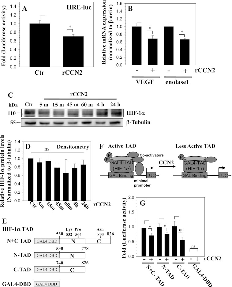
CCN2 treatment decreases the activity of HIF-1α in nucleus pulposus cells. A, cells were transfected with a HIF-1 responsive luciferase reporter (HRE-luc) and activity measured following treatment with 100 ng/ml of rCCN2. CCN2 treatment significantly decreases HRE reporter activity. B, real time RT-PCR analysis of VEGF and ENOLASE 1 mRNA expression in cells treated with rCCN2. Both VEGF and ENOLASE 1 expression decreases with rCCN2 treatment. C, Western blot of HIF-1α in cells treated with rCCN2 for 5 min to 24 h. HIF-1α levels were largely unaffected by the treatment. D, densitometric analysis of three independent experiments in C shows that rCCN2 treatment does not significantly affect HIF-1α levels. E, schematic of TAD constructs used for transactivation studies of HIF-1α. F, schematic summarizing the role of CCN2 in controlling HIF-1α-TAD activity. G, cells were transfected with a Gal4 binary reporter system consisting of Gal4-HIF1α-TAD (HIF-1α N+C TAD, amino acids 530–826; N-TAD, amino acids 530–778; and C-TAD, amino acids 740–826) and pFR-Luc reporter and luciferase activity measured following rCCN2 treatment. Gal4DBD (empty vector pM) was used to measure background pFR-Luc activity. rCCN2 decreased the reporter activity indicating a decreased ability of all HIF-1α-TAD fusion proteins to recruit transcriptional co-activators. Background activity of Gal4DBD was almost undetectable and remained unaffected by CCN2 treatment. Values shown are mean ± S.E. from three independent experiments, *, p < 0.05.

