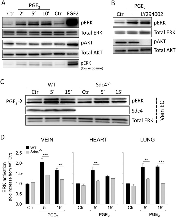
FIGURE 1.
Sdc4 regulates PGE2-induced ERK activation. A, confluent HUVEC were serum-starved and then treated with PGE2 for the indicated times to assess ERK activation (pERK) and AKT activation (pAKT). Ctr, control. B, HUVEC were preincubated with the PI3K inhibitor LY294002 (50 μm) for 30 min before PGE2 stimulation for 5 min. C, primary mouse EC were isolated from WT or Sdc4−/− mice as described under “Experimental Procedures.” Confluent endothelial cells derived from the vein were serum-starved and then treated with PGE2 for the indicated times. Shown is one representative blot of three with similar results. D, pERK quantification in WT versus Sdc4−/− following treatment with PGE2 in EC isolated from different tissues (vein, lung, and heart). Each diagram is derived from three independent experiments (n = 3). Bars represent mean ± S.E. **, p < 0.01; ***, p < 0.001.

