FIGURE 4.
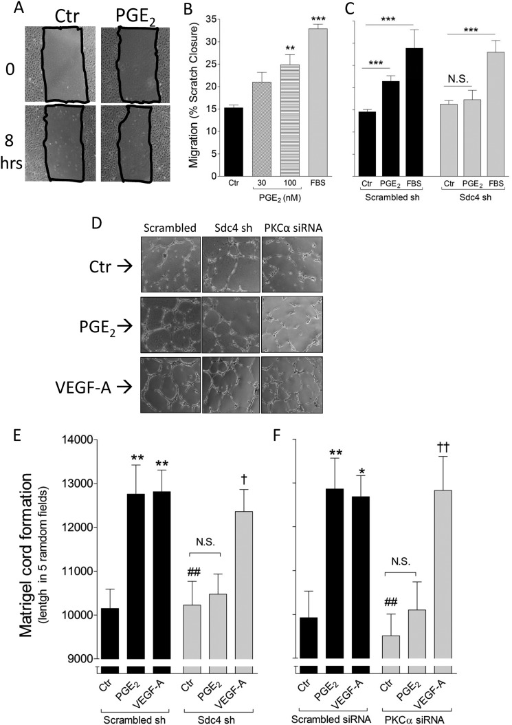
Sdc4 mediates PGE2-induced proangiogenic effect in EC. A, representative pictures of scratch closure in HUVEC. Ctr, control. B, increasing PGE2 concentrations were tested for the ability to induce HUVEC migration. 20% FBS was used as a positive control. The bars represent mean of three independent experiments, each with a quantification of five scratched areas/group of treatment. **, p < 0.01; ***, p < 0.001. C, Scrambled sh or Sdc4 sh HUVEC were treated with PGE2 (100 nm) for 8 h, and the scratch closure was quantified. ***, p < 0.001; N.S., not significant. D, representative pictures of two-dimensional cord formation in the indicated cells in presence of PGE2 or VEGF-A (100 ng/ml). E, in vitro cord formation induced by PGE2 or VEGF-A in Scrambled versus Sdc4 sh HUVEC. The bars represent mean ± S.E. (n = 3). **, p < 0.01 from Scrambled sh Ctr; ##, p < 0.01 from Scrambled sh PGE2; †, p < 0.05 from Sdc4 sh Ctr. F, in vitro cord formation induced by PGE2 or VEGF-A (100 ng/ml) in Scrambled versus PKCα siRNA HUVEC. The bars represent mean ± S.E. (n = 3). *, p < 0.05; **, p < 0.01 from Scrambled siRNA Ctr; ##, p < 0.01 from Scrambled siRNA PGE2; ††, p < 0.01 from PKCα siRNA Ctr).

