FIGURE 1.
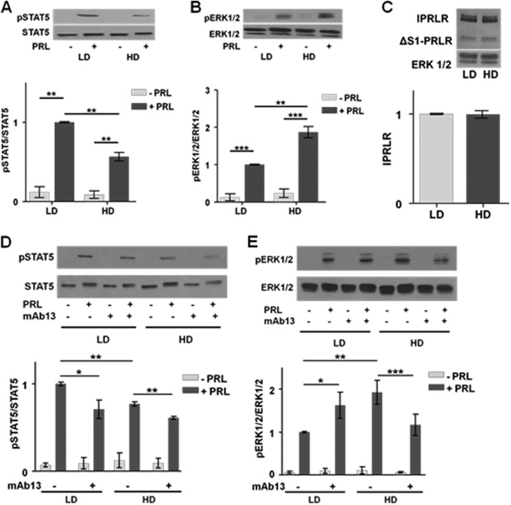
Stiff matrices increase PRL signaling to ERK1/2 and reduce that to STAT5 without altering PRLR levels. A and B, serum-starved T47D cells in LD or HD collagen I gels were treated with or without PRL (4 nm) for 20 min. Cell lysates were immunoblotted with the indicated antibodies. Top panels, representative immunoblots. Bottom panels, quantification of immunoblots by densitometry. Means ± S.D. are shown. n = 3 (A); n = 4 (B). The asterisks denote significant differences between treatments as determined by two-way ANOVA followed by paired t tests. **, p < 0.01; ***, p < 0.001. C, T47D cells in LD or HD collagen I gels were harvested after 24 h of serum starvation, and cell lysates were immunoblotted for PRLR or ERK1/2. Top panel, representative immunoblot. Bottom panel, quantification of the long PRLR isoform (lPRLR) compared with total ERK1/2 by densitometry. Means ± S.D. are shown. n = 3. D and E, serum-starved T47D cells in LD or HD collagen I gels were treated with isotype control antibody (−) or β1 integrin-blocking antibody mAb13 (+) during plating and subsequent treatments as in A and B. Cell lysates were immunoblotted with the indicated antibodies. Top panels, representative immunoblots. Bottom panels, quantification of immunoblots by densitometry. Means ± S.D. are shown. n = 3 (D); n = 4 (E). The asterisks denote significant differences between treatments as determined by two-way ANOVA followed by paired t tests. *, p < 0.05; **, p < 0.01; ***, p < 0.001. Error bars represent S.D.

