FIGURE 3.
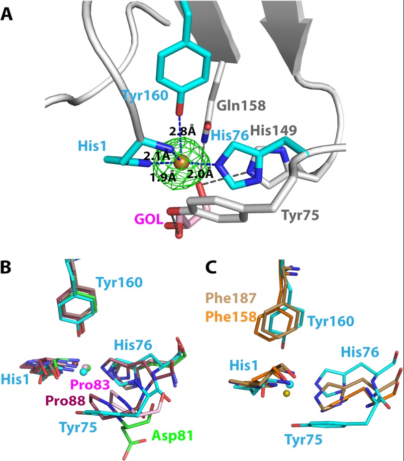
The metal binding site of LPMOs. A, close up view of the PchGH61D in the vicinity of the copper binding site (PDB code 4B5Q). The green Fo − Fc map of the copper atom is contoured at 0.41 e/Å3 (3σ). Cyan-colored residues are coordinated to the copper atom. A glycerol molecule was modeled below the copper atom at the active site, colored in pink (denoted GOL). The bound glycerol molecule is stabilized by His-149, Gln-158, and Tyr-75 (in gray) by hydrogen bonds. B, superposition of the metal binding sites of PchGH61D (PDB code 4B5Q; cyan) with the metal binding sites of NcrPMO-2 (4EIR; green), TauGH61A (3ZUD; pink), and HjeGH61B (2VTC; maroon). The metal ions were modeled as Cu2+ in the first three structures and as Ni2+ in the HjeGH61B structure. C, comparison of the metal binding site of PchGH61D (cyan) with the corresponding non-occupied metal binding sites of SmaCBP21 (2BEM; brown) and EfaCBM33 (4A02; orange).

