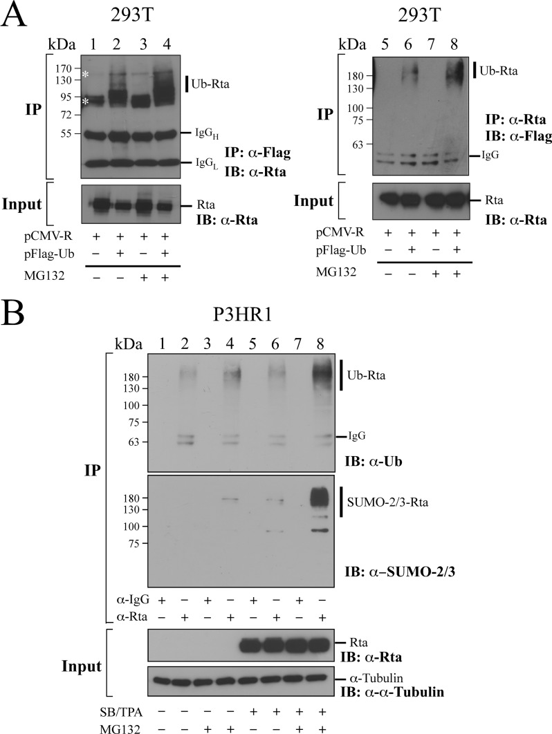
FIGURE 3.
Ubiquitination of Rta in vivo. A, 293T cells were transfected with pCMV-R (lanes 1, 3, 5, and 7) or cotransfected with pCMV-R and pFLAG-Ub (lanes 2, 4, 6, and 8). Cells were treated (lanes 3, 4, 7, and 8) or untreated (lanes 1, 2, 5, and 6) with MG132 for 12 h after transfection for 24 h. Proteins in the cell lysate was immunoprecipitated (IP) using anti-FLAG antibody (lanes 1–4) or anti-Rta antibody (lanes 5–8) after denaturing the proteins in the lysates at 95 °C. The proteins were then detected by immunoblotting (IB) using anti-Rta antibody (lanes 1–4) or anti-FLAG antibody (lanes 5–8). B, P3HR1 cells were treated (lanes 5, 6, 7, and 8) or untreated (lanes 1, 2, 3, and 4) with sodium butyrate and TPA. After incubation for 24 h, cells were treated with MG132 (lanes 3, 4, 7, and 8) or DMSO (lanes 1, 2, 5, and 6) for 12 h. Proteins in cell lysates were immunoprecipitated using anti-Rta antibody and detected by immunoblot analysis with anti-SUMO-2/3 antibody, anti-ubiquitin (Ub), and anti-Rta antibody. Anti-IgG (lanes 1, 3, 5, and 7) was used in IP as a negative control. Asterisks indicate nonspecific bands. Ub-Rta, ubiquitinated Rta; IgGH, the heavy chain of IgG; IgGL, the light chain of IgG.

