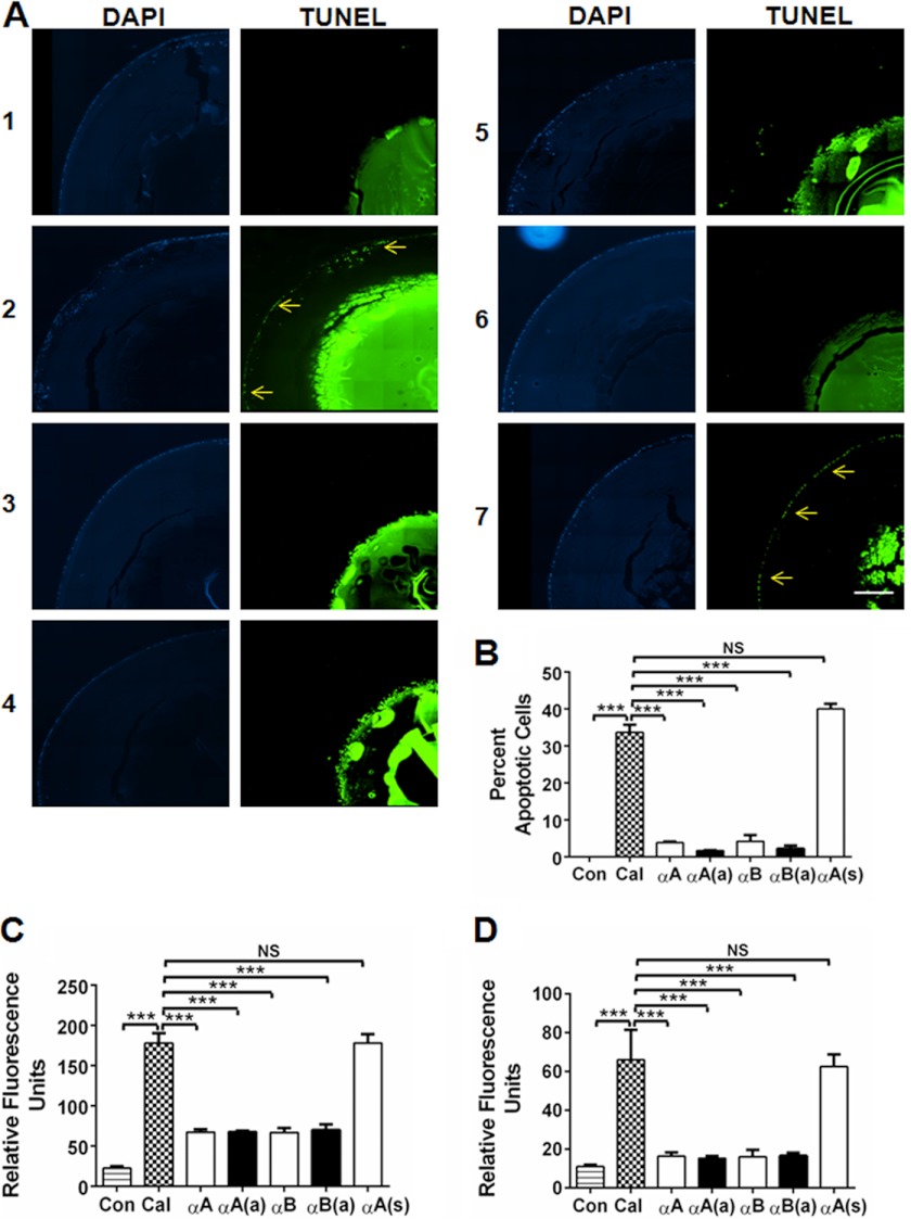
FIGURE 4.
α-Crystallin peptides inhibit calcimycin-induced apoptosis in cultured mouse lenses. Mouse lenses were organ cultured and treated with peptides, as described under “Experimental Procedures.” After treatment, the lenses were thoroughly rinsed in PBS, fixed, and sectioned. The sections were stained to detect apoptotic cells using an in situ Apoptosis Detection Kit (A). Left panels, DAPI staining to visualize the nuclei in the lens epithelium; right panels, TUNEL staining to show apoptosis (arrows). Panel 1, control; panels 2–7, calcimycin-treated lenses. Panel 2, no peptide; panel 3, +αA-native peptide; panel 4, +αA-acetyl peptide; panel 5, +αB peptide; panel 6, +αB-acetyl peptide; and panel 7, scrambled αA-peptide. The percentages of apoptotic cells are presented in a bar graph format in panel B. After culturing, the lenses were homogenized, and caspase-3 (C) as well as caspase-9 (D) activity was determined as described in the legend to Fig. 3. The bars represent the mean ± S.D. of three independent experiments. αA, αA-native peptide; αA(a), αA-acetyl peptide; αB, αB-native peptide; αB(a), αB-acetyl peptide; and αA(s), αA-scrambled peptide. The differences between the native and acetyl peptides were not statistically significant. The intense TUNEL staining in the nuclear region is likely due to fragmented DNA in the terminally differentiated fiber cells. ***, p < 0.0005. NS, not significant. Scale bar = 100 μm.

