FIGURE 9.
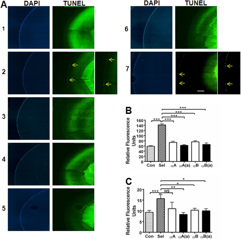
α-Crystallin peptides inhibit apoptosis through inhibition of caspases in lenses with selenite-induced cataract. The lenses from sodium selenite- and sodium selenite + peptide-treated rat pups (multiple 10-μg peptide injections, as in Fig. 5) were fixed and assessed for epithelial cell apoptosis using the In situ Cell Death Detection Kit (panel A). DAPI staining is shown in the left panels, and TUNEL staining is shown on the right. 1, control; lanes 2–6, sodium selenite treated; 2, no peptide, arrows indicate apoptotic cells; 3, +αA-native peptide; 4, +αA-acetyl peptide; 5, +αB peptide; 6, +αB-acetyl peptide; and 7, +αA scrambled peptide. The enlarged images for panel A-2 and A-7 are to show apoptotic cells. The elevation of caspase-3 and caspase-9 activity observed in selenite-induced cataracts was inhibited by both the native and acetyl αA- and αB-crystallin peptides (panels B and C). The bars represent the mean ± S.D. of three independent experiments. αA, αA-native peptide; αA(a), αA-acetyl peptide; αB, αB-native peptide; and αB(a), αB-acetyl peptide. The intense TUNEL staining in the nuclear region is likely due to the reason stated in the legend to Fig. 4. The differences between the native and acetyl peptides were not statistically significant. *, p < 0.05; **, p < 0.005; ***, p < 0.0005. NS, not significant. Scale bar = 100 μm.

