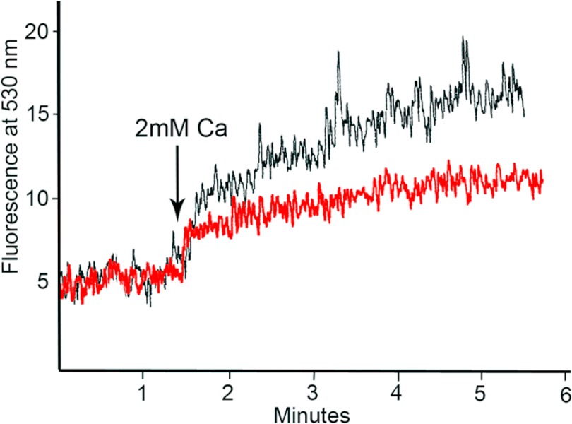
FIGURE 9.
Calcium uptake in the absence of ionophore stimulation. Calcium uptake in RUB1 cells is shown. [Ca 2+]i was measured as described under “Experimental Procedures.” RUB1 cells were loaded with Fluo-3/AM, washed, and suspended in calcium- and magnesium-free flux buffer and then stimulated with 2 mm calcium at 1.5 min. The black trace shows calcium influx by cells treated with nonspecific IgG, whereas the red trace shows calcium influx by cells treated with anti-DPP.

