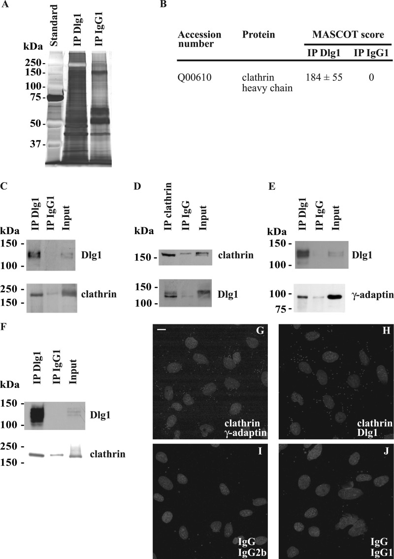
FIGURE 3.
Identification of the coated vesicle components, clathrin heavy chain, and the γ-adaptin subunit of AP-1 as putative direct or indirect Dlg1-interacting partners. A, comparative patterns of the proteins immunoprecipitated from whole hCMEC/D3 lysates using monoclonal Dlg1 antibody (IP Dlg1) or a nonspecific immunoglobulin IgG1 (IP IgG1). Proteins were separated on an SDS-polyacrylamide gel and stained with silver nitrate. B, the proteins immunoprecipitated as mentioned above were trypsinized and identified by tandem mass spectrometry. Identified proteins with a MASCOT score higher than 45 in the IP Dlg1 sample and with a null score in the IP IgG1 sample are considered as putative Dlg1 partners. MASCOT scores are the means ± S.E. from three different experiments. C and D, the presence of clathrin heavy chain in Dlg1 immunoprecipitates was validated by Western blotting. Lysates derived from hCMEC/D3 were immunoprecipitated using a monoclonal Dlg1 antibody (C), a polyclonal clathrin heavy chain antibody (D), or a control IgG. Immunoprecipitates were subjected to Western blotting with Dlg1 or clathrin heavy chain-specific antibodies. E, AP-1 was tested as a Dlg1-interacting partner. Lysates were immunoprecipitated using a polyclonal Dlg1 antibody, and immunoprecipitates were subjected to Western blotting with monoclonal Dlg1 or γ-adaptin antibodies. F, lysates derived from subconfluent Caco-2 cells were immunoprecipitated using a monoclonal Dlg1 antibody or a control IgG1, and immunoprecipitates were subjected to Western blotting with clathrin heavy chain-specific antibodies. The input lane represents 3% of lysate used in immunoprecipitation reactions. G–J, Duolink® PLA labeling of fixed hCMEC/D3 using a polyclonal clathrin heavy chain antibody and a monoclonal γ-adaptin antibody (G) as a positive control to verify the in situ PLA procedure and a monoclonal Dlg1 antibody (H) or, as negative controls, an irrelevant rabbit immunoglobulin IgG and an irrelevant mouse immunoglobulin IgG2b (I) and irrelevant mouse immunoglobulin IgG1 (J). Small gray dots represent the PLA-positive signal. Nuclei appear as gray ellipsoidal organelles. An experiment representative of three independent ones is shown. Scale bar = 10 μm.

