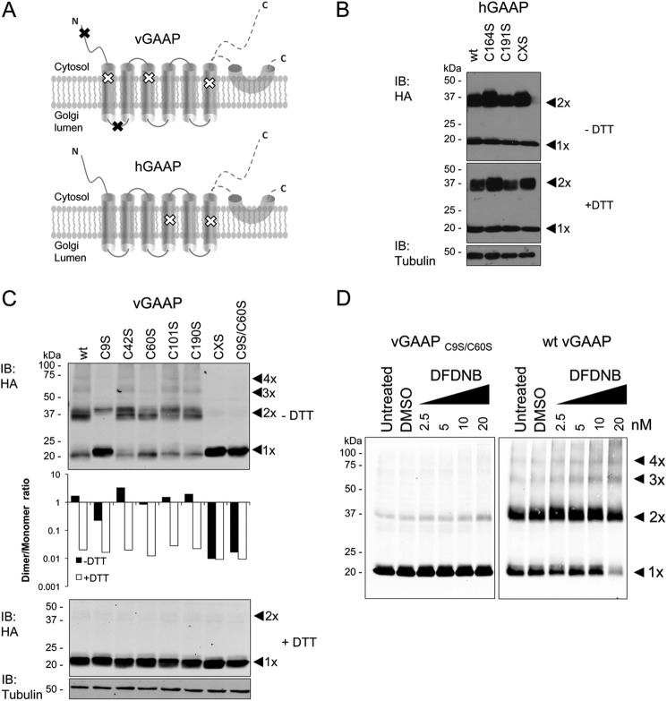
FIGURE 6.
Oligomerization of vGAAP requires cysteine residues 9 and 60. A, distribution of cysteine residues in vGAAP and hGAAP. Black and white x symbols represent cysteine residues that are (black) or are not (white) needed for oligomerization of vGAAP. GAAP topology was described previously (9), where the region after TMD6 was suggested to be either a re-entrant loop or cytosolic. Cysteine to serine mutations were made in hGAAP-HA (B) or vGAAP-HA (C). Lysates from HeLa cells transfected with wild type or mutant vGAAP-HA or hGAAP-HA were analyzed by SDS-PAGE with or without DTT and immunoblotted (IB) with anti-HA antibody. C, top panel: dimer and monomer band intensities of the vGAAP-HA mutants were measured using Li-COR and plotted as the ratio between dimer and monomer (C, middle panel). CXS denotes mutants with all cysteines mutated to serines. D, lysates from HeLa cells transfected with wild type or C9S/C60S vGAAP-HA were incubated with DFDNB, resolved by non-reducing SDS-PAGE, and immunoblotted with anti-HA antibody. The predicted oligomeric states of vGAAP are shown by arrowheads. Results shown are representative of three independent experiments. The sizes of molecular mass markers are shown in kDa. DMSO, dimethyl sulfoxide.

