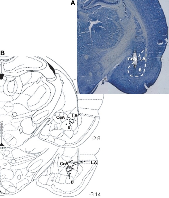
Figure 1.

(A) Photomicrograph illustrating the positioning of a stimulating electrode tip in BLA. Brain section at about 3.14 mm posterior to bregma. (B) A diagram depicting a coronal section of the rat brain (at position 3.14 mm posterior to bregma) showing the location of the stimulating electrode tip in the BLA, for an average pool of 30 animals (LA, lateral amygdala; B, basal amygdala; CeA, central amygdala). Diagram is from the stereotaxic atlas of Paxinos and Watson (1997).
