Figure 4.
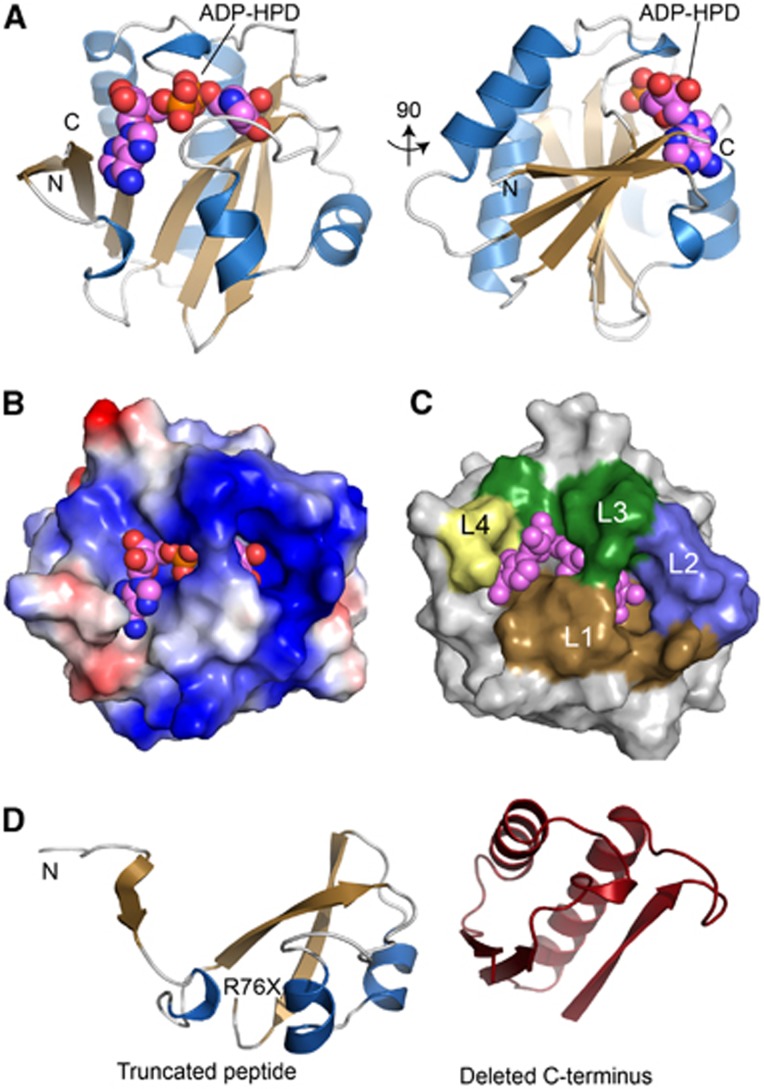
Crystal structure of the C6orf130 ADP-HPD complex. (A) Orthogonal views of C6orf130 (blue and tan) bound to ADP-HPD (spheres). (B) Surface charge representation of C6orf130 (blue, positive; red, negative; grey, neutral or hydrophobic) showing a positively charged binding site for ADP-HPD. (C) ADP-HPD binding site is composed of four sequence motifs, L1–L4. (D) The C6orf130 protein truncation (R76X) in the patients analysed in this study.

