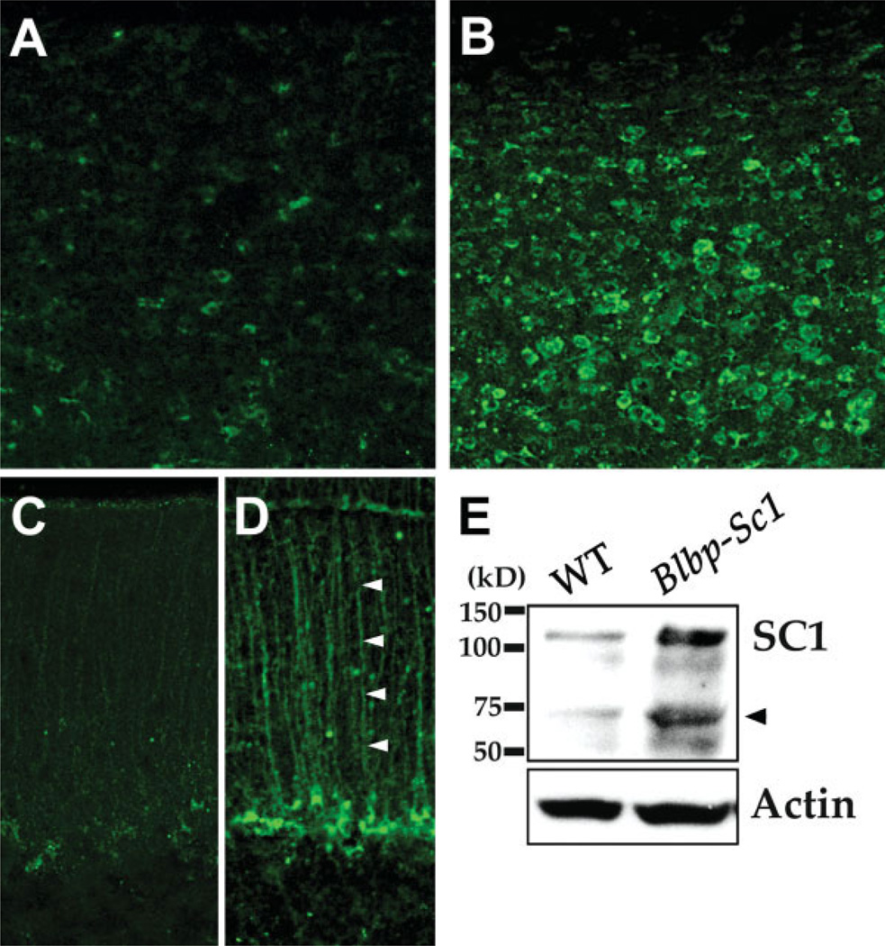
Fig. 3.
Elevated expression of SC1 in the cerebral cortex and cerebellum of Blbp-Sc1 transgenic mice sections of adult cortex and cerebellum were immunolabeled with anti-SC1 antibodies. Increased levels of SC1 are seen in astroglial cells of Blbp-Sc1 transgenic mice within both the cortex (B) and the cerebellum (D), as compared to basal levels of SC1 expression in wildtype mice (A [cortex], C [cerebellum]). Arrowheads (D) indicate Bergmann glia of the cerebellum. Immunoblot analysis of Blbp-Sc1 brains demonstrates an increase in the levels of the 116 kDa SC1 protein (E). Arrowhead (E) indicates the unprocessed precursor of SC1. [Color figure can be viewed in the online issue, which is available at www.interscience.wiley.com.]

