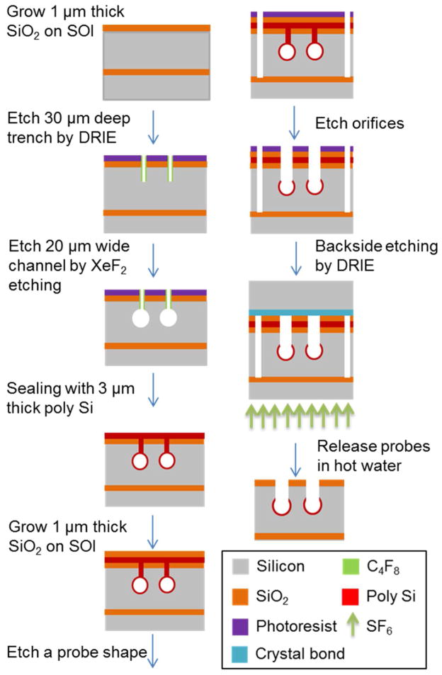
Figure 2.
Sequential steps for microfabrication of Si probe. Device is shown in cross-section. 20 μm wide channels were formed using Xactix XeF2 (isotropic etching) through 30 μm deep trenches and sealed with poly silicon. Deep reactive ion etching (DRIE) then cut probe shapes and sampling orifices. Probes were thinned by the backside etching by DRIE on a silicon support and released in hot water. See Experimental in supporting information for details.

